3D images of leaves: a photo shoot we should all be looking at
In our modern age of “plant blindness” – where people underappreciate the plants around us – Professor Margaret Barbour and her PhD student, Richard Harwood, are imaging leaves in three dimensions. Not only will their findings further our understanding of how leaves function, these images could help us to recognise just how vital plants are to life on Earth
Plants could arguably be said to be the unsung champions of life as we know it. Rare is the day that we do not encounter a plant in some form or another, from walking by hedgerows on our way to school to sitting at the dinner table with the family to eat. Indeed, plants underpin all life on Earth, including humans. The air we breathe and the food we eat comes from plant life, and yet scientists still have a lot to learn about plants.
Based at the School of Life and Environmental Sciences, University of Sydney, Professor Margaret Barbour and her PhD student, Richard Harwood, are imaging leaves in three dimensions. By imaging leaves in this way, Margaret and Richard are hoping to develop our understanding of leaf function and answer questions that have eluded scientists so far.
QUICK FACTS
ORGANELLES are tiny structures inside cells that work together to perform specific tasks.
CHLOROPLASTS (shown right in green in the 3D image of a chickpea mesophyll cell) are organelles found in green algae and plant cells. Their job is to help turn sunlight into food that can be used by the cell, through a process called photosynthesis.
MITOCHONDRIA (shown right in red) are organelles that exist in the cells of plants and animals. Often called the “powerhouses of the cell”, their function is to break down sugars and create energy- carrier molecules for the cell.
VACUOLES are also organelles found in animal and plant cells. Filled with fluid, vacuoles are the space in the middle of the cell and have many important functions, including storing nutrients and waste products to help cells survive. There is usually one large vacuole in each plant cell.
HOW DOES THE 3D IMAGING OF LEAVES WORK?
Most of our knowledge of leaf anatomy and how this links to leaf function comes from 2D cross-sections of leaves. However, the leaf interior is a complex 3D arrangement of cells and tissues that allow light capture and gas (oxygen and carbon dioxide) diffusion for photosynthesis, as well as regulation of water transport. Margaret and Richard have developed a microscopic technique that, for the first time, enables 3D imaging of leaf cells and their organelles, including chloroplasts and mitochondria.
WHAT TECHNOLOGY IS INVOLVED IN CREATING THE 3D IMAGES?
Margaret and Richard take a very small sample of a leaf, replacing all of the water in the leaf sample with a chemical solution called glutaraldehyde under a gentle vacuum to fix all the structures in place. They then use heavy metal staining to enhance the contrast between different structures, such as cell walls and chloroplast membranes, and to compare these with the large vacuole in the middle of the cells. The sample is then embedded in resin and trimmed to make sure they are imaging the correct part of the leaf.
“Imaging is carried out using a field emission scanning electron microscope that has an automatic microtome [a tool for cutting extremely thin slices],” explains Margaret. “An image of the surface of the sample is taken, then the microtome cuts a very thin slice off the top and another image is taken. This is repeated 800 times to build up a stack of images that can be analysed using 3D reconstruction software.”
DID THE 3D IMAGING TECHNOLOGY ALREADY EXIST?
The field emission scanning electron microscope has been widely used for animal samples like liver and kidney tissues. Plant tissue is harder to image because plant cell walls require special sample preparation. In the first instances, the team spent a long time optimising the fixing, staining and embedding steps before they managed to get good images. They still need to optimise the sample preparation for every new species they image in 3D because plant cell walls and the density of cells in leaves vary a lot between species.
WHAT DOES 3D IMAGING TECHNOLOGY ENABLE US TO LEARN?
One of the first things Margaret and Richard learned is that mitochondria are shaped like worms, not round or kidney bean-shaped like the drawings in textbooks. In plant cells, the mitochondria are pressed right up against chloroplasts, and a single mitochondrion can be both adjacent to the cell wall and on the interior side of a chloroplast at different positions along its length. All current models of photosynthesis and respiration assume that mitochondria are in one position or the other – not both.
Estimating the surface area to volume ratio is important because it enables us to better understand how it relates to photosynthesis; plants need to balance their need for more surface area to collect sunlight, with the fragility of their leaves and the rate of water loss. Thanks to 3D imaging technology, scientists now know that simple 2D geometric shapes, like spheres and capsules, do not accurately estimate the surface area and volume of leaf cells and chloroplasts.
TAKE A LOOK INSIDE A CHICKPEA PLANT LEAF!
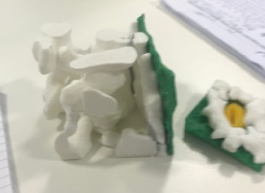
Stomata are tiny openings or pores in the plant tissue that allow plants to “breathe”, i.e. carbon dioxide enters, and water and oxygen exit, through a leaf’s stomata. You can take a look inside – and even print – a chickpea stomata in four easy steps.
1) Download these four chickpea stomata .stl files.
2) Drag and drop any one of these files into this free STL viewer: https://www.viewstl.com.
3) This STL viewer will allow you to zoom in and out, and spin the part of the stomata you have chosen around.
4) If your school or college has access to a 3D printer, you can have a go at printing a 3D image of a chickpea stomata. In Windows 10, double click on one of the files you have downloaded and it will open in Print 3D. This is what your stomata will look like when it is printed!
Cuticle.stl | Epidermal Cells.stl | Guard Cells.stl | Spongy Mesophyll Cells.stl
WHAT ARE THE BENEFITS OF BEING ABLE TO LOOK AT LEAVES IN THIS WAY?
With authentic 3D anatomical models of cells and organelles, the researchers will be able to predict important leaf functions like photosynthesis, respiration and transpiration (when plants absorb water through the roots and release water vapour through pores in the leaves) more accurately. They will also be able to run experiments on different types of leaf anatomies that allow higher rates of photosynthesis, which is good news for agriculture. Scientists could use the information to genetically modify crops to grow faster and use water more efficiently. “We have genetic tools to alter leaf anatomy, but transforming crop plants is time consuming and expensive,” says Margaret. “So, if we can predict the effects before genetic modification, we can save time and money.”
Reference
https://doi.org/10.33424/FUTURUM29

3D image of a chickpea mesophyll cell
© Richard Harwood
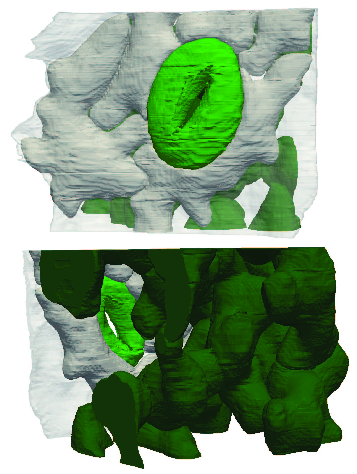
3D reconstruction of a chickpea stoma (tiny pores in the epidermis of the leaf or stem of a plant, shown in pale green), epidermal cells (white) and mesophyll cells (dark green)
© Richard Harwood and Quinn Musulin
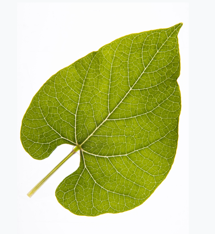
Common bean (Phaseolus vulgaris) leaf
© William Salter
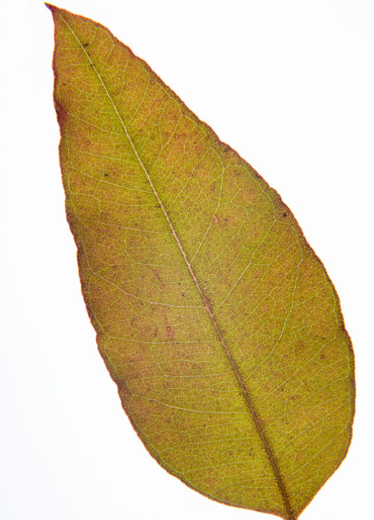
Parramatta Red Gum (Eucalyptus parramattensis) leaf
© William Salter

Adopt a plant! Succulents are hardy, but you’ll need to be patient because they grow really
slowly.
© Margaret Barbour
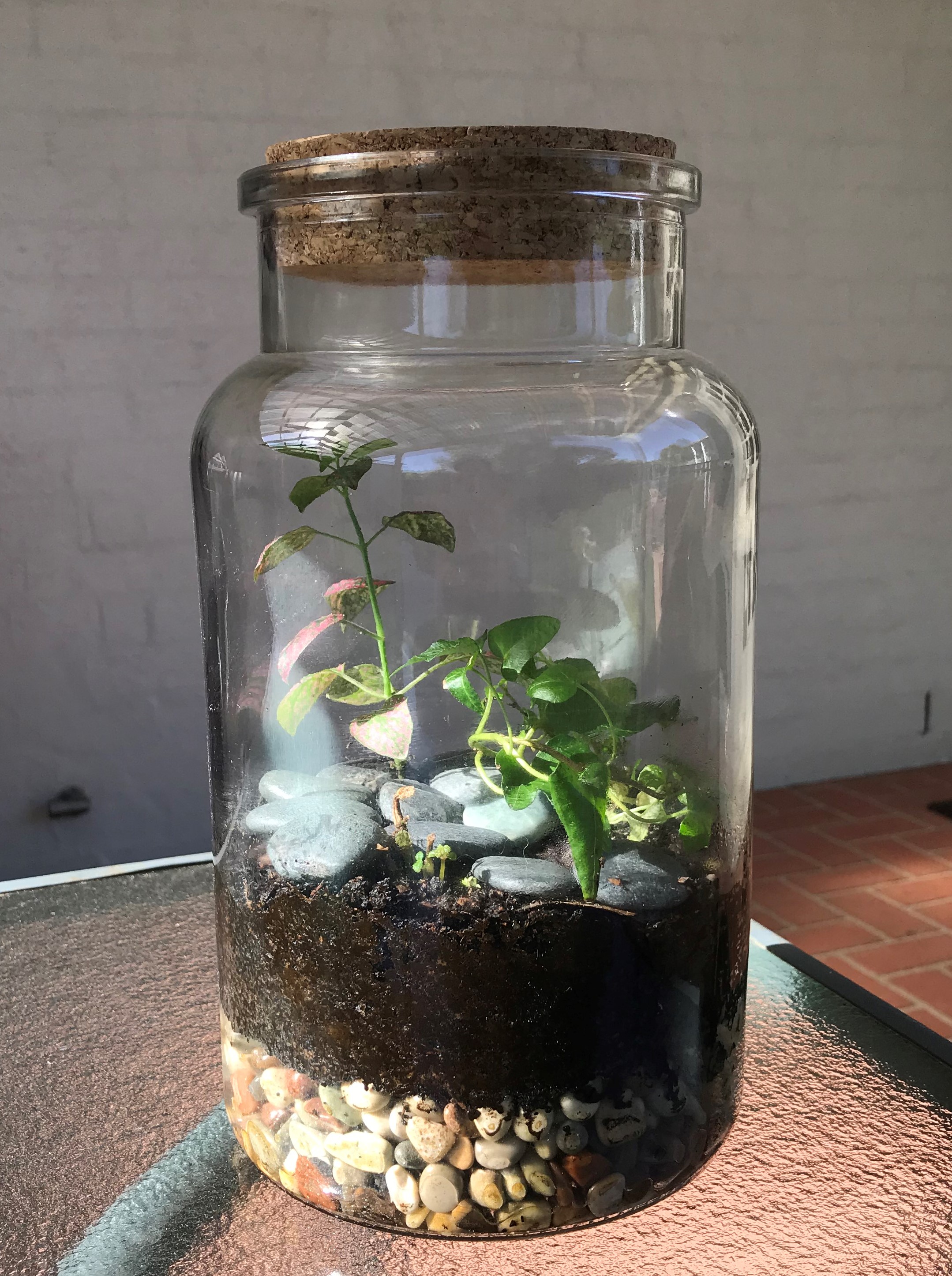
Terraria are fun to make, and you can watch ecosystem water and carbon cycles in miniature.
© Margaret Barbour
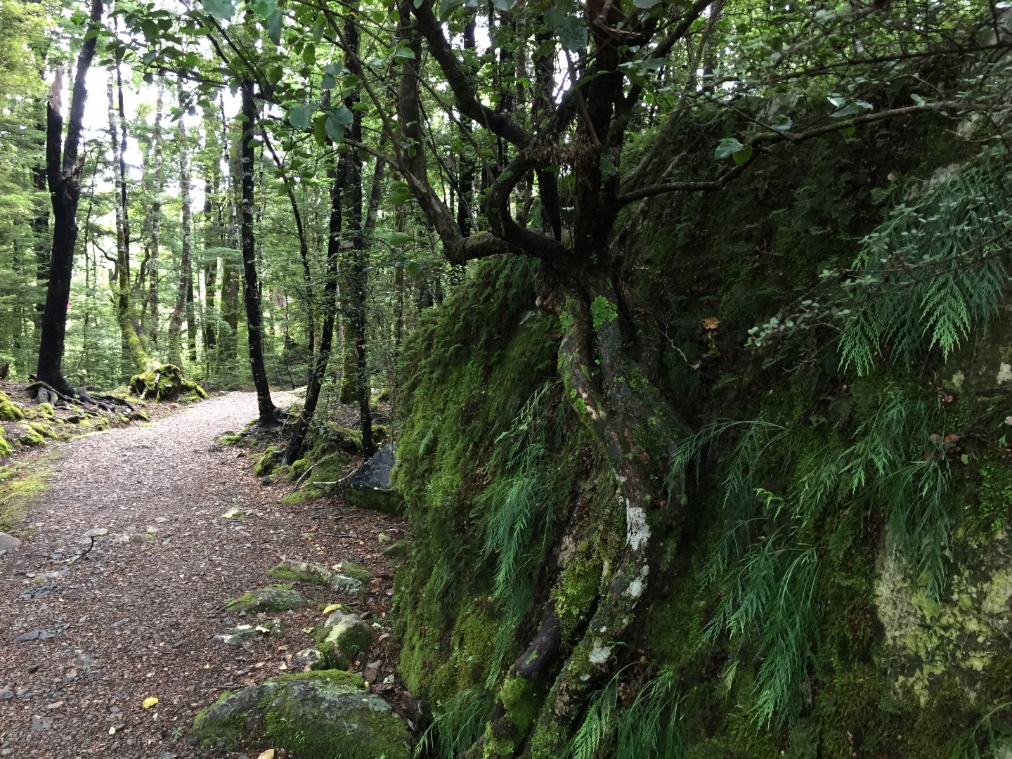
Southern Beech (Nothofagus solandri) forest in the South Island of New Zealand
© Margaret Barbour
Incredibly, people tend to ignore or underappreciate plants despite their importance to humans and all life on Earth; this is called “plant blindness”. Margaret and Richard hope that by creating a virtual reality experience of the inside of leaves, people will be able to step out of their human-centric view of life and into the plant world.
WHAT QUESTIONS WILL THESE NEW IMAGING TECHNIQUES ENABLE US TO ANSWER?
There are currently major gaps in our understanding of how leaves work. For example, it is still not known where water changes phase from liquid to gas. This is important because leaves are a major part of the global water cycle – between 50% and 70% of rainfall returns to the atmosphere via leaves. It is known that there is a large resistance to the diffusion of CO2 within leaves – and that it limits photosynthesis – but nobody knows exactly where it is yet. If this resistance could be reduced, we could increase photosynthesis and therefore crop yield.
Of course, developing a fuller understanding of plant mechanisms is important in its own right, but with a burgeoning global population and climate change, the need to find ways to improve crop productivity and identify how plants respond to future climates is ever- increasing.
Plants could arguably be said to be the unsung champions of life as we know it. Rare is the day that we do not encounter a plant in some form or another, from walking by hedgerows on our way to school to sitting at the dinner table with the family to eat. Indeed, plants underpin all life on Earth, including humans. The air we breathe and the food we eat comes from plant life, and yet scientists still have a lot to learn about plants.
Based at the School of Life and Environmental Sciences, University of Sydney, Professor Margaret Barbour and her PhD student, Richard Harwood, are imaging leaves in three dimensions. By imaging leaves in this way, Margaret and Richard are hoping to develop our understanding of leaf function and answer questions that have eluded scientists so far.
QUICK FACTS
ORGANELLES are tiny structures inside cells that work together to perform specific tasks.
CHLOROPLASTS (shown right in green in the 3D image of a chickpea mesophyll cell) are organelles found in green algae and plant cells. Their job is to help turn sunlight into food that can be used by the cell, through a process called photosynthesis.
MITOCHONDRIA (shown right in red) are organelles that exist in the cells of plants and animals. Often called the “powerhouses of the cell”, their function is to break down sugars and create energy- carrier molecules for the cell.
VACUOLES are also organelles found in animal and plant cells. Filled with fluid, vacuoles are the space in the middle of the cell and have many important functions, including storing nutrients and waste products to help cells survive. There is usually one large vacuole in each plant cell.
HOW DOES THE 3D IMAGING OF LEAVES WORK?
Most of our knowledge of leaf anatomy and how this links to leaf function comes from 2D cross-sections of leaves. However, the leaf interior is a complex 3D arrangement of cells and tissues that allow light capture and gas (oxygen and carbon dioxide) diffusion for photosynthesis, as well as regulation of water transport. Margaret and Richard have developed a microscopic technique that, for the first time, enables 3D imaging of leaf cells and their organelles, including chloroplasts and mitochondria.
WHAT TECHNOLOGY IS INVOLVED IN CREATING THE 3D IMAGES?
Margaret and Richard take a very small sample of a leaf, replacing all of the water in the leaf sample with a chemical solution called glutaraldehyde under a gentle vacuum to fix all the structures in place. They then use heavy metal staining to enhance the contrast between different structures, such as cell walls and chloroplast membranes, and to compare these with the large vacuole in the middle of the cells. The sample is then embedded in resin and trimmed to make sure they are imaging the correct part of the leaf.
“Imaging is carried out using a field emission scanning electron microscope that has an automatic microtome [a tool for cutting extremely thin slices],” explains Margaret. “An image of the surface of the sample is taken, then the microtome cuts a very thin slice off the top and another image is taken. This is repeated 800 times to build up a stack of images that can be analysed using 3D reconstruction software.”
DID THE 3D IMAGING TECHNOLOGY ALREADY EXIST?
The field emission scanning electron microscope has been widely used for animal samples like liver and kidney tissues. Plant tissue is harder to image because plant cell walls require special sample preparation. In the first instances, the team spent a long time optimising the fixing, staining and embedding steps before they managed to get good images. They still need to optimise the sample preparation for every new species they image in 3D because plant cell walls and the density of cells in leaves vary a lot between species.
WHAT DOES 3D IMAGING TECHNOLOGY ENABLE US TO LEARN?
One of the first things Margaret and Richard learned is that mitochondria are shaped like worms, not round or kidney bean-shaped like the drawings in textbooks. In plant cells, the mitochondria are pressed right up against chloroplasts, and a single mitochondrion can be both adjacent to the cell wall and on the interior side of a chloroplast at different positions along its length. All current models of photosynthesis and respiration assume that mitochondria are in one position or the other – not both.
Estimating the surface area to volume ratio is important because it enables us to better understand how it relates to photosynthesis; plants need to balance their need for more surface area to collect sunlight, with the fragility of their leaves and the rate of water loss. Thanks to 3D imaging technology, scientists now know that simple 2D geometric shapes, like spheres and capsules, do not accurately estimate the surface area and volume of leaf cells and chloroplasts.
TAKE A LOOK INSIDE A CHICKPEA PLANT LEAF!
Stomata are tiny openings or pores in the plant tissue that allow plants to “breathe”, i.e. carbon dioxide enters, and water and oxygen exit, through a leaf’s stomata. You can take a look inside – and even print – a chickpea stomata in four easy steps.
1) Download these four chickpea stomata .stl files.
2) Drag and drop any one of these files into this free STL viewer: https://www.viewstl.com.
3) This STL viewer will allow you to zoom in and out, and spin the part of the stomata you have chosen around.
4) If your school or college has access to a 3D printer, you can have a go at printing a 3D image of a chickpea stomata. In Windows 10, double click on one of the files you have downloaded and it will open in Print 3D. This is what your stomata will look like when it is printed!
Cuticle.stl | Epidermal Cells.stl | Guard Cells.stl | Spongy Mesophyll Cells.stl
WHAT ARE THE BENEFITS OF BEING ABLE TO LOOK AT LEAVES IN THIS WAY?
With authentic 3D anatomical models of cells and organelles, the researchers will be able to predict important leaf functions like photosynthesis, respiration and transpiration (when plants absorb water through the roots and release water vapour through pores in the leaves) more accurately. They will also be able to run experiments on different types of leaf anatomies that allow higher rates of photosynthesis, which is good news for agriculture. Scientists could use the information to genetically modify crops to grow faster and use water more efficiently. “We have genetic tools to alter leaf anatomy, but transforming crop plants is time consuming and expensive,” says Margaret. “So, if we can predict the effects before genetic modification, we can save time and money.”
Incredibly, people tend to ignore or underappreciate plants despite their importance to humans and all life on Earth; this is called “plant blindness”. Margaret and Richard hope that by creating a virtual reality experience of the inside of leaves, people will be able to step out of their human-centric view of life and into the plant world.
WHAT QUESTIONS WILL THESE NEW IMAGING TECHNIQUES ENABLE US TO ANSWER?
There are currently major gaps in our understanding of how leaves work. For example, it is still not known where water changes phase from liquid to gas. This is important because leaves are a major part of the global water cycle – between 50% and 70% of rainfall returns to the atmosphere via leaves. It is known that there is a large resistance to the diffusion of CO2 within leaves – and that it limits photosynthesis – but nobody knows exactly where it is yet. If this resistance could be reduced, we could increase photosynthesis and therefore crop yield.
Of course, developing a fuller understanding of plant mechanisms is important in its own right, but with a burgeoning global population and climate change, the need to find ways to improve crop productivity and identify how plants respond to future climates is ever- increasing.
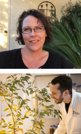 MARGARET BARBOUR
Professor of Plant Physiology, School of Life and Environmental Sciences, University of Sydney, Australia
RICHARD HARWOOD
PhD student, School of Life and Environmental Sciences University of Sydney, Australia
FIELD OF RESEARCH: Plant Physiology
RESEARCH PROJECT: Professor Margaret Barbour and Richard Harwood are investigating a means of imaging leaves in three dimensions. The findings will lead to increased understanding of leaf functions, such as photosynthesis and the regulation of water transport
FUNDERS: Australian Research Council, Grains Research and Development Corporation, University of Sydney Strategic Research Excellence Initiative
MARGARET BARBOUR
Professor of Plant Physiology School of Life and Environmental Sciences University of Sydney, Australia
RICHARD HARWOOD
PhD student
School of Life and Environmental Sciences University of Sydney, Australia
FIELD OF RESEARCH
Plant Physiology
RESEARCH PROJECT
Professor Margaret Barbour and Richard Harwood are investigating a means of imaging leaves in three dimensions. The findings will lead to increased understanding of leaf functions, such as photosynthesis and the regulation of water transport
FUNDERS
Australian Research Council, Grains Research and Development Corporation, University of Sydney Strategic Research Excellence Initiative
ABOUT PLANT SCIENCE
Plant science goes by many different names – botany, plant biology or phytology – but they all essentially mean the same thing, which is the science of plant life. It should be understood as a branch (no pun intended) of biology, which has evolved over time to become a broad, multidisciplinary subject involving a range of other areas of science and technology.
HOW HAS PLANT SCIENCE EVOLVED OVER THE YEARS?
When people first began studying plants, their main concerns were describing what they looked like, where they grew, what they could be used for and how related they were to one another. Sometime during the 17th century, however, scientists started to turn their investigations towards understanding how plants function – how they grow, how they sense and respond to their environment, their biochemical pathways and, more recently, the molecular genetics of plants. Today, all of these topics of interest fit within the broad area of plant physiology.
WHERE DID THE IDEA TO IMAGE LEAVES IN 3D STEM FROM?
When scientists first saw the structure of a leaf through microscopes, they recognised that there was a complicated 3D structure and organisation within leaves. They built beautiful wax models of the inside of leaves to represent what they saw and teach botany students about plants. You can still see some very early wax models of leaves at the Orta Botanico di Pisa botanical garden.
“Our team has wanted to image the inside of leaves in 3D ever since we saw 3D X-ray computed tomography images of soil and roots in the mid-2000’s,” explains Margaret. “We recognised just how powerful 3D images would be to advance our understanding of leaf function.”
THE ROYAL SOCIETY OF BIOLOGY SAYS THERE IS A SHORTAGE OF PLANT SCIENTISTS IN THE UK. CAN THE SAME BE SAID FOR AUSTRALIA?
Yes. There is a shortage of plant scientists in the agriculture industry, in biosecurity and in biodiversity conservation in Australia. Problems associated with global warming and climate change will only increase the need for plant scientists in Australia and around the world, so the field is ripe for students who are interested in developing a career in the field.
THE SOCIETY ALSO CLAIMS THAT PLANT SCIENTISTS ARE RESPONDING TO SOME OF THE MOST CRITICAL CHALLENGES OF THE 21ST CENTURY. WHAT ARE THESE CHALLENGES?
Some the most pressing issues facing humanity require an understanding of how plants function, including understanding and responding to climate change, biodiversity conservation, addressing food and nutrition security, and planning our cities for the future. In fact, of the 17 UN sustainable development goals (SDGs), plant science contributes to seven of them. The contribution that plant scientists can make to an ever-changing world can hardly be overstated.
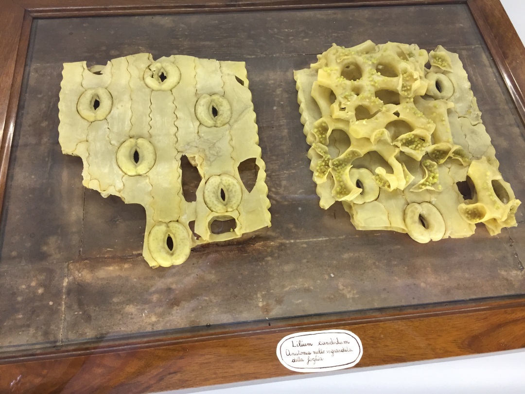
An anatomical model of the surface and inside of a lily leaf on display at the Orta Botanico di Pisa, Italy.
© Margaret Barbour
OPPORTUNITIES IN PLANT SCIENCE
• There are almost unlimited career opportunities in the field of plant science. Options include agronomist, biochemist, entomologist, horticulturist, soil scientist and toxicologist, forestry researcher, gardening show TV host – and options will only increase in the future! Just take a look at CropLife International.
• According to PayScale, the average salary for a botanist in Australia is AU$68,837
• The Chartered Institute of Horticulture has loads of information about how you can make a difference by studying the field of plant science and technology
ASK PROF MARGARET BARBOUR AND RICHARD HARWOOD
WHAT DID YOU WANT TO BE WHEN YOU WERE YOUNGER?
MB: I wanted to be a scientist from when I was around 8 years old.
RH: It changed from day to day when I was a child, but I have always been pretty optimistic! Becoming a pro surfer was one of my more ambitious plans, especially considering I lived 200km from the coast.
WHO OR WHAT INSPIRED YOU TO GET INTO PLANT SCIENCE?
MB: My parents gave me a little microscope for my eighth birthday and showed me how to take an epidermal peal from a Daphne plant growing outside our home. I saw stomata for the first time and realised that plants were so much more active than I had imagined – they were sensing and responding to the world in really cool ways. I was hooked on plants from then on.
RH: I decided to study environmental science at university because I always enjoyed being out in nature. The course I studied offered field trips to amazing places around Australia and that sealed the deal! Being blown away by how much carbon and water is constantly moving around plants is what inspired me to embark on a plant science research degree.
WHAT DO YOU LOVE MOST ABOUT YOUR WORK?
MB: I love stretching my mind to think about plants and the environment in new ways. It is very satisfying to come up with novel measurement techniques and theoretical models to test an aspect of plant function, and then be able to make those ideas happen.
RH: I love merging research and technology. A highlight of my PhD has been learning about virtual reality and 3D printing.
DOES WORKING WITH TECHNOLOGY SUCH AS 3D IMAGING STOP YOU FROM WORKING IN NATURAL ENVIRONMENTS?
MB: If I’m honest it does keep me indoors more than I’d like. I need to make more time for walking in forests and touching leaves!
RH: It does – our 3D imaging work has me glued to the computer screen most days. Fortunately, my desk overlooks some beautiful trees for when I need a quick nature fix.
FROM A PLANT SCIENCE PERSPECTIVE, WHAT WOULD YOU SAY ARE THE MOST PRESSING CHALLENGES OF THE 21ST CENTURY?
MB: I think the big challenges in plant science are: a) predicting how future climates will impact natural ecosystems; b) adapting our crops to secure our food and nutrition; c) improving crop productivity and water-use efficiency; and d) changing the way humans value plants.
RH: I think plant science will play a pivotal role in dealing with climate change and sustainably feeding a growing population. A particular challenge is developing better communication between scientists and policymakers to address these issues in an effective way.
MARGARET’S TOP TIPS
01 – Study as many maths, physics and chemistry-related subjects as you can at school. Even if these subjects don’t seem very relevant to biology when you are in school, they will be really helpful later on.
02 – Grow some plants at home! It’s amazing how much you can learn just by looking closely at a living houseplant.
03 – Visit your local botanic gardens – most have great displays on plant evolution. Ask the people in charge of looking after them plenty of questions – their insight will prove invaluable.
RICHARD’S TOP TIPS
01 – My number one tip is to keep in mind how diverse plant science is. Plant scientists can be found in labs and on computers, but they can also be found 30 metres up in the air in the jungle or shaking snow off leaves in tundra!