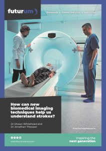
How can new biomedical imaging techniques help us understand strokes?
Strokes can be devastating. Depending on their severity, they can cause issues with movement and muscle control, a loss of feeling and sensation, and degenerative diseases such as dementia. Understanding how strokes affect the brain can help researchers develop therapeutic interventions that aid recovery. Dr Jonathan Thiessen and Dr Shawn Whitehead from Western University, in Canada, are studying how to combine positron emission tomography (PET) and magnetic resonance imaging (MRI) to gain a better understanding of the effects of strokes and how to treat them.
Talk like a biomedical imaging specialist
Biomarker — a biological molecule or characteristic that aids the diagnosis of disease
Dementia — a generic term describing impaired ability to think, remember or make decisions. Alzheimer’s disease is the most common type of dementia
Imaging — the process of making a visual representation of something by scanning it with a detector and processing the results
Magnetic resonance imaging (MRI) — a non-invasive biomedical imaging technology that uses magnetic and radio waves to produce three-dimensional images of certain structures in the body
Positron emission tomography (PET) — a minimally-invasive biomedical imaging technology that uses positrons to produce three-dimensional images of certain structures in the body
Positron — a positively charged particle considered the ‘antiparticle’ of the electron, as it has the same mass and magnitude of charge
Stroke — a medical condition that happens when blood supply to part of the brain is restricted or cut off
When they are working as they should, our brains are nothing short of miraculous. Within the gooey, pink lump that sits inside your skull, billions of neurons are communicating via tiny sparks of electricity, keeping you alive and allowing you to interact with the world around you.
Unfortunately, a healthy brain is not a privilege shared by everyone. Just like cars and computers, as our brains get older, they become more susceptible to the wear and tear of daily use. The older we get, the more likely we are to suffer from health conditions such as stroke and dementia.
One in three people over the age of 60 will have a stroke, develop dementia or suffer from both during their lifetime. “A stroke happens when insufficient blood is reaching brain tissue,” says Dr Shawn Whitehead from Western University. “This means that the oxygen, glucose and other nutrients that the brain needs do not arrive on time.” This can cause a loss of neurons, damage to brain blood vessels and increased inflammation and swelling of the brain. “Depending on the severity and location of the stroke, outcomes can include loss of motor function, loss of sensation and even cognitive impairment and dementia,” says Shawn.
Shawn and his colleague Dr Jonathan Thiessen are investigating exactly how strokes affect the brain, and how this links to the likelihood of a patient developing dementia at a later stage. To do this, they are using two biomedical imaging technologies, combining the images produced by both to understand the complex interactions happening inside the brain.
PET and MRI
Two biomedical imaging technologies stand out as the most useful for examining the living brain’s functions and structure. “Positron emission tomography (PET) can measure tiny traces of molecules, drugs or cells that have been labelled with a radioactive atom that emits particles called positrons,” says Jonathan. “When a positron meets an electron, they create a burst of energy that we can detect with PET.”
The labelled molecules are called PET tracers, and they have no effect on the body’s functions because they are only used in tiny amounts. “PET tracers can measure blood flow, metabolism and types of brain cells,” says Jonathan. “These measurements can provide unique insights into how the brain’s functions change after a stroke.”
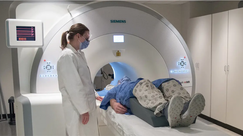
Magnetic resonance imaging (MRI) works in a different way. “Hydrogen atoms in water molecules have a property called ‘spin’ that aligns with the direction of a magnetic field,” explains Jonathan. “MRI uses a powerful magnet to align the spins of hydrogen atoms in the body, before using pulses of radio energy to knock the hydrogen atoms out of equilibrium.” After the pulse passes, the atoms return to equilibrium and can be measured and imaged. Because so much of the body is made up of water, the resultant images can highlight differences between healthy and diseased tissues, including in the brain. “MRI can detect early signs of stroke, assess stroke severity and type, and assess levels of recovery,” says Jonathan.
Combining PET and MRI
PET and MRI provide different insights about the body, but it is when they are used together that they are most powerful. “It’s possible to acquire PET and MRI images at exactly the same time,” says Jonathan. “This means that the resulting images are perfectly aligned in space and time.”
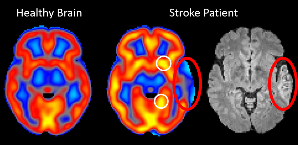
While MRI images lend insights into brain tissue structure and chemistry, PET images tell us about levels of inflammation in the brain, the presence of abnormal proteins associated with dementia and the density of connections between neurons. “The sum result of PET/MRI systems is truly greater than its parts,” says Jonathan. “This is why we are developing PET/MRI techniques that detect changes in the brain after a stroke.” This is important because there is evidence that leaving such changes unaddressed, even if they do not appear to have any immediate effect on a person’s abilities, can lead to the early onset of dementia.
From research to practice
Shawn and Jonathan hope that their findings will lead to new therapeutic practices that could be used in hospitals and clinics to help recovering stroke patients. Research suggests that strokes can increase the number of clumps of abnormal proteins, known as amyloid plaques, found in the brain. Such plaques can be a precursor to dementia and bring about the degeneration of neuronal pathways and overall cognitive decline. Clinicians use the presence of these proteins in the blood as a biomarker – a biological sign that suggests dementia is present or likely to develop.
Biomarkers alone, however, do not tell researchers how the brain is functioning after a stroke. “The benefit of PET/MRI imaging is that we can get a sense of how the brain looks and functions prior to, and at different stages after, a stroke,” explains Shawn. “In combination with bloodbased biomarkers, we can develop an approach to determine who is at risk for developing dementia after a stroke.” Once clinicians have this information, they can recommend therapeutic approaches for patients to help their recovery and minimise the chances of dementia developing.
 Dr Jonathan Thiessen
Associate Professor, Department of Medical Biophysics and Department of Medical Imaging, Schulich School of Medicine and Dentistry, Western University, Canada
Dr Shawn Whitehead
Associate Professor, Department of Anatomy and Cell Biology, Schulich School of Medicine and Dentistry, Western University, Canada
Field of research: Biomedical imaging
Research project: Combining PET and MRI imaging technologies to deepen understanding of the effects of strokes on the brain
Funders: Natural Sciences and Engineering Research Council (NSERC), Canada Foundation for Innovation, Ontario Research Fund, Mitacs, Canadian Institutes of Health Research, New Frontiers in Research Fund, Ontario Institute of Cancer Research, Lawson Health Research Institute, Western University
Reference
https://doi.org/10.33424/FUTURUM504
A. PET images showing inflammation in the brain following a stroke (orange blob). B. PET images showing the influence of exercise following a stroke on the density of connections between neurons. Exercise may help improve brain function after a stroke.
About biomedical imaging
Biomedical imaging involves creating images of biological tissues and processes as they happen, in living people or other organisms. X-rays and ultrasounds are well-known examples. Such technologies are hugely useful for researchers and clinicians, as they allow them to see what is happening inside the body without the need for surgery.
“Biomedical imaging is always advancing,” says Jonathan. “New tools are improving our ability to see small features, decrease the time needed to take images and enhance our ability to analyse different tissue properties.” The combination of diagnostic imaging with targeted therapeutics, known as ‘theranostics’, is helping clinicians identify and treat diseases in real time, and see the effects of treatment on the body in more detail.
Advances in positron emission tomography (PET) technologies include whole-body PET systems, that can image the entire body at once, and head-only PET systems, that hone in on small features of the brain. “MRI is also constantly progressing,” says Shawn. “Larger magnetic fields are improving image quality and portable MRI systems are making the technology more accessible.” MRI equipment is typically large and expensive, and only functions in specially magnetically-shielded rooms. Overcoming these limitations is helping biomedical imaging to become feasible in remote and under-resourced areas of the world.
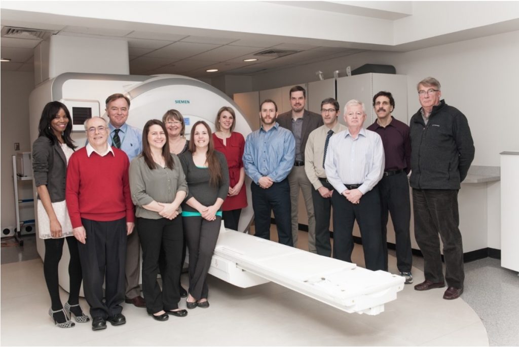
These processes involve a lot of people with different specialties working together. “There are many steps involved in developing a new imaging method or PET tracer, determining which brain tissues or functions it best images and demonstrating its usefulness,” explains Jonathan. “Once that’s all in place, the next step is to translate it into clinical research and practice.” Such an effort inevitably requires collaboration from many scientists and clinicians covering a wide range of expertise, as well as trainees, such as graduate or postgraduate students, and a range of technicians who oversee the development, operation and maintenance of the equipment itself.
Pathway from school to biomedical imaging
At school, relevant subjects include physics, chemistry, mathematics, computer science and biology.
Jonathan recommends seeking out experts in the areas that interest you and asking questions about their career paths. Securing work experience in a lab or hospital that deals with biomedical imaging technologies would also provide you with an insight into what careers in the field involve.
Jonathan initially studied physics at university, which gave him a good understanding of how imaging techniques work at the molecular and subatomic level. Other relevant courses include medical biophysics, biomedical engineering and computer science. Many other courses, such as medicine, neuroscience, psychology and biology may also be useful.
Jonathan also notes vocational routes outside of university studies, including training to become a radiologist, medical physicist or medical radiation technologist.
Explore careers in biomedical imaging
The Schulich School of Medicine and Dentistry at Western University runs an outreach programme called Experiential Learning Academy for Biomedical Sciences (X-LABS) that introduces high school students to hands-on research experience.
There are a variety of societies that cover areas related to biomedical imaging technologies, which provide a range of learning resources and opportunities to connect with people in the field. These include the International Society for Magnetic Resonance in Medicine, the Society of Nuclear Medicine and Molecular Imaging and the World Molecular Imaging Society.
Opportunities in biomedical imaging span academia, clinical care and industry. Salaries also vary accordingly. For example, according to Glassdoor, the average salary for a Medical Imaging Technologist in Canada is around CAN $69.7k a year.
Meet Shawn
As a kid, I wanted to grow up to be a bus driver. I think I always wanted to do something that would help people.
I love working with talented people. I work with incredibly smart students and postdocs, which keeps me creative in my work. Seeing the work of our lab receive increasing recognition has been a massive highlight.
My lab uses an interdisciplinary approach to screen for dementia following stroke. We use a combination of animal behaviour, imaging, blood-based biomarkers and molecular biology approaches. Much of my day is spent overseeing the research done by postdocs and graduate students, as well as liaising with experts like Jonathan. I also lead Western’s Neuroscience Institute. Going forward, I want to develop screening tools for dementia that are as accessible as possible.
I don’t have much free time to unwind. Four kids keep me very busy outside of work!
Shawn’s top tip
Don’t try to copy anyone else’s career path. Do something that drives you to get out of bed in the morning and make a difference.
Meet Jonathan
I grew up on a farm in rural Manitoba. Though I was always interested in science and technology, I didn’t have a clear career path until I was well into university. The most important step for me was taking on a degree and discovering all the possibilities that are out there.
I studied physics as an undergraduate and graduate student. My first exposure to biomedical imaging was as a summer research student during my undergraduate degree, where we used MRI to study Alzheimer’s disease. I was instantly hooked by the ‘magic’ of seeing the brain non-invasively and focused on MRI for my PhD. After completing my PhD, I pivoted to PET as a postdoctoral fellow, before moving to study both MRI and PET in combination.
I love working with brilliant students and colleagues. As well as imaging the brain, I collaborate with clinicians and scientists using PET/MRI to better understand and treat cancer, infection, cardiac disease and musculoskeletal conditions. I am Director of the PET/MRI Facility, which means much of my day is spent interacting with scientists and trainees, but there will also be time dedicated to reading, writing and editing grants or journal articles, and teaching.
Between work and three kids, I’m kept pretty busy. I try to find time at the end of the day to read a good book or watch a funny show. My spouse and I go for a walk every evening, and seek out hiking trails on the weekends to spend time in nature.
Jonathan’s top tip
Pursue a career that is interesting and challenging for you personally. Seek out experts in the area you’re interested in and get their advice. Remember, everyone’s path is different, and that’s OK.
Do you have a question for Shawn and Jonathan?
Write it in the comments box below and Shawn and Jonathan will get back to you. (Remember, researchers are very busy people, so you may have to wait a few days.)
Discover more about new biomedical imaging techniques and how they work:
www.futurumcareers.com/how-does-physics-allow-us-to-look-inside-the-body





0 Comments