How does physics allow us to look inside the body?
Biomedical imaging techniques enable doctors and scientists to view inside the human body, allowing them to diagnose medical conditions and uncover the biological processes that are essential for life. At the University of California Davis, USA, Professor Simon Cherry and Professor Ramsey Badawi have developed the world’s first full-body scanner, which has the potential to revolutionise medicine and biomedical research.
Talk like a biomedical imaging specialist
Magnetic resonance imaging (MRI) — a biomedical imaging technique that uses magnets and radio waves to observe internal organs
Metabolism — the chemical reactions in cells that convert food into energy
Photon — a particle of light
Positron — a positively charged subatomic particle with the same mass as an electron
Positron emission tomography (PET) — a biomedical imaging technique that detects signals emitted from radiotracers injected into the body to measure biological functions
Radioactive decay — the emission of energy in the form of ionising radiation
Radioisotope — an unstable form of an element that undergoes radioactive decay
Radiotracer — a very small (trace) amount of a chemical compound to which a radioisotope has been attached
What do you think of when you hear words like ‘positron’, ‘antimatter’ and ‘high-energy photon’? Physics is full of exotic-sounding terms that we might associate with science fiction, and even real physicists, such as Albert Einstein and Erwin Schrödinger, can take on surreal and almost mythical personas. It can be easy to forget that these words equate to real-life phenomena, and these scientists made discoveries that have practical implications in our daily lives.
Every day, in hospitals all over the world, doctors use the laws of physics to diagnose and treat their patients. X-ray machines, ultrasound devices, magnetic resonance imaging (MRI) scanners and positron emission tomography (PET) scanners were all developed using concepts and theories based in physics. These techniques capture internal images of parts of the body, allowing medical professionals to diagnose a wide range of health conditions, from broken bones to cancer.
At the University of California Davis, Professor Simon Cherry and Professor Ramsey Badawi have taken biomedical imaging one step further. They have developed the EXPLORER total-body PET scanner, the world’s first biomedical imaging device that can capture an image of the entire body in a single snapshot.
What physics theories lie behind PET scanners?
“Before having a PET scan, the patient is injected with a tiny amount of radiotracer,” explains Simon. “One common example is a sugar molecule that has been modified to contain the radioisotope fluorine-18.” After injection, the radiotracer circulates throughout the body and is taken up by cells. The more metabolically active the cells are, the more radioactive sugar they take up. This is diagnostically useful because, during certain diseases, the body changes how it uses sugars. For example, cancer cells often accumulate much more radioactive sugar than surrounding tissues, meaning they can be visually detected on a PET scan. The major clinical application of PET is to look for cancer cells in patients.
About an hour after the patient has been injected with the radiotracer (allowing time for it to be distributed throughout the body), they are placed inside a PET scanner, which can detect the signals coming from radioactive decay. “When a fluorine-18 atom in the radiotracer undergoes radioactive decay, it emits a positron,” explains Ramsey. “Positrons are antiparticles of electrons, meaning radioactive decay produces antimatter!”
However, matter and antimatter cannot coexist for very long. Almost immediately, the positron will find a nearby electron and both particles will be annihilated. In the process, the positron and electron disappear, and their mass is converted into energy according to Einstein’s famous E = mc2 equation.
This energy is released as two high-energy photons emitted in opposite directions. The radiation detectors in the PET scanner look for this signature pair of photons arriving simultaneously on opposite sides of the body, indicating that radioactive decay has occurred. During a single PET scan, hundreds of millions of decay signals will be detected, which reveal where radioactive molecules are concentrated in the body, indicating the location of cancer cells or other abnormalities.
How does EXPLORER differ from conventional PET scanners?
The EXPLORER scanner is an incredibly complex machine with over half a million radiation detectors and more than 50,000 electronic channels. “This rivals the complexity of the detectors used in the big particle accelerators found at places like CERN, home of the Large Hadron Collider!” says Simon.
Conventional PET scanners have a small array of radiation detectors and can only image part of the body at a time. To study the whole body, a patient is moved through the scanner step by step (which is time-consuming) and then the individual scans must be ‘stitched’ together. In contrast, EXPLORER has many more radiation detectors that cover the entire length of the body. This allows it to capture images of the whole body in a single scan – the world’s first biomedical imaging device to do so.
How does EXPLORER benefit patients and biomedical researchers?
With detectors covering the whole length of the body, EXPLORER can detect radiation much more efficiently, gathering a lot more information. For a given scan time or radiation dosage, EXPLORER can collect 40 times more signal than a conventional PET scanner, providing many benefits for patients and biomedical researchers.
For example, EXPLORER can collect higher quality images which could enable diseases to be diagnosed earlier. It can conduct scans 40 times faster, meaning a total-body scan can be completed in less than two minutes. “This is fantastic for patients who can’t stay still for long periods of time, such as young children or patients in a lot of pain,” says Ramsey. Or, for a standard scan time, the patient can be injected with a radioactive dosage that is 40 times lower. “This is great for patients who need regular PET scans.”
“But the real game changer,” says Simon, “is that EXPLORER can see the radiotracer wherever it is in the body at any moment in time.” This allows scientists to observe how substances are transported around the body, delivered to tissues and metabolised by cells, providing insights into how the entire body functions. “This offers truly exciting prospects for the future!” says Ramsey.
How did Simon and Ramsey develop EXPLORER?
In addition to the technical challenges of developing such a complex machine, Simon and Ramsey also had to overcome the practical challenges that come with creating a new invention, in particular, the issue of money. “It was a 15-year journey from first ideas to first images,” says Ramsey. “The first 10 years were very frustrating, as the idea of a total-body scanner didn’t interest the biomedical community, so we couldn’t get any funding.”
This all changed in 2015, when Simon and Ramsey finally received a large grant from the NIH, providing the money necessary to start building the system. In 2016, they joined forces with United Imaging Healthcare, a medical imaging company with the physical resources to build the scanner. “After that, the project moved incredibly quickly,” says Ramsey. “Our first full-body images were acquired in 2018, and we started using EXPLORER for clinical use in 2019. Now, there are almost 20 EXPLORER total-body PET scanners around the world, which have scanned well over 100,000 people. Other companies also are making similar scanners, which shows that the idea is now catching on.”
“Looking back, it has been an exciting and worthwhile journey,” says Simon. The team’s dedication and persistence paid off, and thousands of people are now benefitting from the technology Simon and Ramsey have developed. Having improved the diagnosis of cancer, Simon and Ramsey envision that, in future, total-body PET will be combined with other advanced techniques (for example, genomics and proteomics) and artificial intelligence to predict how the human body responds to health challenges and diseases, revolutionising biomedical understanding.
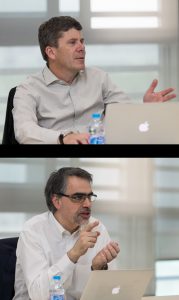 Professor Simon Cherry
Professor Ramsey Badawi
Department of Biomedical Engineering, University of California Davis, USA
Fields of research: Biomedical imaging, biomedical engineering, medical physics
Research project: Developing EXPLORER – the world’s first total-body biomedical imaging scanner
Website: explorer.ucdavis.edu
Funder: US National Institutes for Health (NIH)

Reference
https://doi.org/10.33424/FUTURUM445


Conventional PET scanners have a small array of radiation detectors and so can only image
part of the body at a time
© Cherry et al., 2017
(www.science.org/doi/10.1126/scitranslmed.aaf6169)

The EXPLORER total-body PET scanner has radiation detectors along the entire length of the body, meaning it can image the whole body at the same time
© Cherry et al., 2017
(www.science.org/doi/10.1126/scitranslmed.aaf6169)

EXPLORER total-body PET scanners are now used in hospitals around the world
About biomedical imaging
How does physics impact medicine?
Biomedical imaging is a good example of how physics impacts medicine, as many techniques use some form of electromagnetic radiation to produce images of the body. At the low energy end of the electromagnetic spectrum, radio waves create the signal for MRI scans, while X-rays and gamma rays (the high-energy photons in PET) are at the opposite end of the spectrum.
“X-rays and gamma rays are ionising radiation, meaning they can break chemical bonds,” explains Ramsey. “Therefore, care is always taken to minimise the radiation dose during X-rays and PET scans.” If you break a bone and have an X-ray, you will receive the equivalent of about 10 days of natural background radiation. If you have a conventional PET scan, you will receive the equivalent of about 8 years of background radiation. One of the advantages of EXPLORER is that, when necessary, it can do PET scans with extremely low radiation doses – equivalent to the radiation you would receive during a round trip flight from San Francisco to London!
In contrast, ultrasound imaging uses soundwaves, which are not part of the electromagnetic spectrum. These soundwaves are of such a high frequency that we cannot hear them. When the soundwaves bounce of different body parts, they create an ‘echo’ which is detected by the ultrasound probe and transformed into a moving image of the body’s interior.
Why do so many different imaging techniques exist?
“Each imaging technique provides different information at different levels of detail and at different costs,” explains Simon. X-rays are perfect for providing images of anatomical issues, such as broken bones and fluid in the lungs. Ultrasound does not expose the patient to any radiation, so is used to generate real-time images of a foetus in the womb. Both these techniques are low cost, so are widely used. On the other hand, MRI and PET scans are more expensive but can provide more detailed images. MRI scans generate 3D images of body parts, especially soft tissues, while PET scans produce functional images of biological processes, such as metabolism or blood flow.
Pathway from school to biomedical imaging
• At school, you will learn about the processes behind biomedical imaging in physics, about human anatomy in biology, and how to use and manipulate equations in maths. If classes are available, computer science and engineering will also be very useful.
• Depending on what area of biomedical imaging you would like to work in, there are different pathways you can take to a career in the field. If you want to design and build biomedical imaging devices, consider a degree in almost any field of engineering (biomedical, mechanical, electrical or even aerospace), biomedical imaging, physics or medical physics. If you want to use biomedical imaging techniques while working with patients, consider a degree in radiography or medicine.
• Look for research opportunities and internships with biomedical research labs to learn about and contribute to scientific advances in biomedical imaging. Most labs have websites where you can learn about their research (Simon’s lab: cherrylab.bme.ucdavis.edu; Ramsey’s lab: badawilab.bme.ucdavis.edu) and many host high school and undergraduate students.
• Look for work experience and job shadowing opportunities with radiographers and medical imaging technicians in your local hospital to learn how biomedical imaging is used in clinical settings
Explore careers in biomedical imaging
• Careers in biomedical imaging are wide-ranging and varied, spanning engineering developments, scientific advances and clinical applications.
• Biomedical engineers and medical physicists, who may work in industrial companies or research institutions, such as universities, develop new biomedical imaging technologies and methods, while chemists develop new radiotracers for PET scanning.
• In hospitals, biomedical imaging devices are operated by radiographers or technologists and maintained by medical technicians and hospital physicists. Doctors, who may specialise in radiology or nuclear medicine, interpret the scans and use them to diagnose and treat patients’ conditions.
• Biomedical researchers use biomedical images to learn more about the human body.
• The National Institute of Biomedical Imaging and Bioengineering has a wealth of resources for students, including fun facts, competitions, games and advice for future scientists: www.nibib.nih.gov/science-education
Meet Simon
I was football crazy as a kid. I was always kicking a ball about, and I loved watching my local team, Sheffield United. I’ve also always enjoyed music, especially classical music. I started piano lessons when I was six and have continued on and off to this day. In my teens, I became interested in astronomy, so I got a telescope and joined a junior astronomical society.
My father was a biophysicist, and I would visit his labs during the holidays. This, combined with my love of astronomy and my great physics and chemistry teachers, gave me a love of science. I never really considered doing anything else for a career, except when I was ten, when I wanted to be a Formula 1 racing driver!
At university, I studied physics with astronomy. I wanted to continue with physics research for a PhD, but I didn’t know which area I wanted to work in. I had never heard of medical physics, but when I discovered it involved the atomic and nuclear physics topics that interested me, combined with very direct practical applications in medical imaging and radiation therapy, I decided to focus on it. I was accepted to work on project studying PET imaging. At the time, I didn’t have a clue what I was getting myself into, but it turned out to be a perfect fit for me.
A willingness to trust and collaborate with others is important for scientists, and I am proud of the interactive, collaborative research environment in biomedical imaging that we have created at the University of California Davis. My success has also stemmed from perseverance and an aptitude for bold, difficult challenges.
I love the fact that with PET imaging, we are using fundamental theories and discoveries of physics. Radioactive decay, antimatter and the conversion of positrons and electrons into photons based on Einstein’s famous E = mc2 equation sounds like science fiction, but we use this every day in hospitals to help patients.
Simon’s top tips
1. Believe in your ideas, even when others do not.
2. Be kind and respectful to those you work with – you will be repaid many times over.
3. Always pay attention to work-life balance. Having interests and time away from research will make you a better researcher.
Meet Ramsey
I was interested in aircraft when I was younger, and I memorised the shapes and designs of many civil and military aeroplanes. As I grew older, I became fascinated by strategy games, and my friends and I developed various ‘world conquest’-style board games. All very nerdy!
I always knew I wanted to be a scientist – strangely enough, from even before I really knew what it meant! In high school, I was good at physics and chemistry, and I had tremendous teachers in those subjects. I loved learning about the Voyager space probes, which sent remarkable images of the planets back to Earth. So, when I had the opportunity to study physics at university, it was a natural choice.
I worked in aerospace software engineering for a while, then got a job as a systems administrator at the Clinical PET Centre at St. Thomas’ Hospital in London. This was the first clinical PET service in the UK. I barely knew what PET was at the time, but it caught my interest and before I knew it, I was doing a part-time PhD in PET imaging!
I have worked with some tremendous young scientists over the years and helping them develop their full potential as researchers has been enormously rewarding. That’s a gift that will keep on giving and will hopefully outlast me and the various projects I have been lucky enough to participate in.
Science is too broad for one person to master. If you want to make an impact, you need to collaborate, so the ability to work with and get on with other people is really important.
I love the way that PET allows you to see fundamental processes of biology, like the metabolism of sugar, happening right before your eyes in the living body. It’s an astonishing trick!
Ramsey’s top tips
1. Believe in your dreams. Listen to the nay-sayers (you might learn something), but don’t let them stop you pursuing your ideas.
2. Have a ‘back-up’ plan. While Simon and I spent 10 years persuading people to fund EXPLORER, I was also working on other projects to pay the bills and keep the lights on in the lab.
3. Always stay in touch with your inner child that jumps up and down with excitement at your latest scientific idea!
Do you have a question for Simon or Ramsey?
Write it in the comments box below and the Simon or Ramsey will get back to you. (Remember, researchers are very busy people, so you may have to wait a few days.)
Hi Goodafternoon Mr. Ramsey:
Becoming a doctor in medicine has been my passion as long as I can remember. I have no idea what I am getting myself involved with please highlight me. My commitment to coordinating, collaborating and cooperating with other medical professionals is one of the reason I would like to pursue medicine.
How do I know what is right for me? career wise. Teaching was a second career choice but two years and change my mind is not there, it does not align with what I really want to study.
Becoming a medical doctor is very challenging, but very rewarding! You need to be willing to memorize lots and lots of facts about biology, anatomy and other topics like physiology. You also need to not get too grossed-out by the sticky, goopy stuff you find inside people! The training process is long, and once you have your MD degree, you are not done – you still have to train to become a specialist, which could take another 4 years or more. But, you get to work with patients, figure out what is wrong with them, and help them recover. What a wonderful thing to do every day! But, don’t underestimate the amount of time and hard work you will have to put in over many years. You won’t have much time for hobbies and such.