Can functional imaging and image-guided therapy revolutionise treatments for neurological disorders?
Functional imaging allows us to see how our bodies work from the inside out, while image-guided therapy uses information from medical images to make treatments more precise, more effective and less invasive. With cutting-edge tools like focused ultrasound and advanced brain imaging, Dr Bojana Stefanovic and Dr Meaghan O’Reilly, researchers at Sunnybrook Research Institute and the University of Toronto in Canada, are developing innovative, non-invasive treatments for neurological disorders.
Talk like a … medical imaging researcher
Alzheimer’s disease — a condition that affects the brain, causing progressive problems with memory, leading to confusion, loss of independence and, ultimately, death
Focused ultrasound — a non-invasive medical treatment that uses targeted ultrasound waves to deposit energy inside the brain
In vivo — a procedure that takes place within a living body
Neurological disorder — a disease or illness that affects the brain, spinal cord or nerves
Spinal cord — the long, tube-like structure that runs from the brain and down the back, transmitting signals between the brain and the rest of the body
Two-photon fluorescence microscopy — an optical imaging technique that provides the most detailed images of the living brain, letting researchers visualise individual brain cells
Ultrasound — high-frequency sound waves, beyond the human range of hearing
Traditionally, many diseases that affect the brain or spinal cord have been treated through surgery. However, these surgical procedures are often invasive and can come with risks and long recovery times, so researchers are now developing non-invasive techniques for operating on the brain. These new techniques allow doctors to target problem areas without making a single incision.
Using functional brain imaging techniques and image-guided therapies, Dr Bojana Stefanovic and Dr Meaghan O’Reilly from Sunnybrook Research Institute and the University of Toronto are developing revolutionary treatments and techniques that could provide precise, non-invasive medical care for some of the most challenging neurological disorders.
What is image-guided therapy?
“Image-guided therapy uses medical imaging to plan and deliver medical interventions,” explains Meaghan. “These image-guided procedures are less invasive and often more effective than traditional surgery.” New brain function imaging techniques can pinpoint damaged regions of the brain, allowing doctors to target these areas during treatment. “With image-guided therapy, treatments can be focused exclusively on the affected regions while sparing surrounding healthy areas,” continues Meaghan. “This reduces the likelihood of side effects such as behavioural or cognitive problems, which are common when healthy brain tissue is harmed.”
“Medical images can also allow doctors to assess the effects of a treatment on brain functioning, sometimes even while a procedure is still taking place,” says Bojana. “For example, doctors can find out in real-time whether they have successfully restored connections between damaged regions of the brain.” This helps doctors make efficient, well-informed decisions about how to proceed with their patient’s treatment.
Looking inside the brain
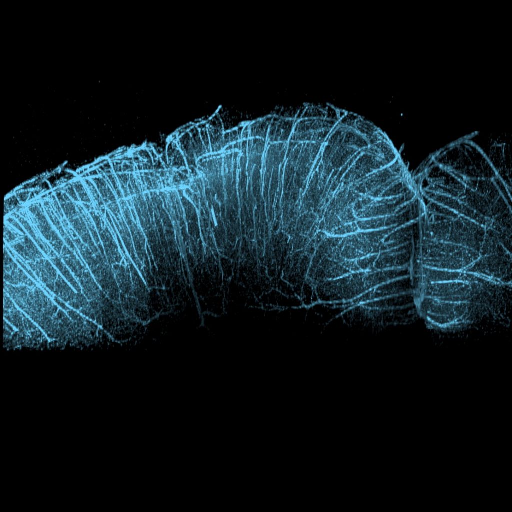
Studying live brain tissue is extremely challenging as light scatters when it passes through the scalp and skull, making it impossible to form clear images of brain tissue at depth. An imaging technique called two-photon fluorescence microscopy overcomes this problem by using near-infrared light, which scatters less than visible light, allowing it to travel deeper into opaque substances, such as the living brain.
Brain cells can be labelled with fluorescent markers, called fluorophores, which emit light when they relax from an excited state brought about by lasers within the microscope. These lasers produce extremely short pulses of high-frequency light which are precisely focused at a tiny volume of the brain, enabling sub-micrometre resolution images of brain cells and their activity. Unlike traditional imaging methods, this in vivo technique allows scientists to study the brain at the cellular level while it is at work.
“Alzheimer’s disease, stroke and brain trauma all affect the functioning and structure of brain cells,” says Bojana. “Two-photon fluorescence microscopy lets us study these changes in great detail, as well as the effects of new interventions, teaching us more about brain diseases and helping us characterise the effects of novel treatments on damaged brain cells.”
The power of sound
Focused ultrasound uses sound waves to treat areas inside the body without the need for incisions. A bowl-shaped device, known as a transducer, vibrates and produces sound waves which are focused towards a point in the middle of the bowl. At this point, called the focus, the energy that was spread out over the whole surface of the bowl becomes concentrated into a small volume. “The energy in that focus can be aimed at target tissue deep in the brain, while the transducer remains outside of the body,” explains Meaghan. “This is incisionless brain surgery!”
Focused ultrasound can be used to treat a range of neurological disorders by adjusting the pressure and duration of the sound waves. “Delivering such focused energy can create permanent effects, such as destroying tumours by heating them, or temporary effects, such as changing the permeability of blood vessels to help drugs reach the brain tissue in higher amounts,” says Meaghan.
The lining of blood vessels prevents harmful substances from entering the brain or spinal cord, but they can also block helpful drugs from reaching their target tissue. “Tiny bubbles, called microbubbles, can be injected into the bloodstream and caused to vibrate using focused ultrasound, increasing the permeability of the blood vessel walls,” explains Meaghan. “For a few hours, drugs that normally would not be able to reach the brain or spinal cord are able to pass out of the blood vessels and reach their targets in the surrounding tissue.” This method is already being tested in patients with brain tumours and Alzheimer’s disease. However, more work needs to be done to develop a tool capable of precisely directing sound waves into the spinal cord.
What does the future hold?
“The spinal cord is both incredibly important and also highly sensitive to injury, so the benefits of a completely non-invasive treatment would be huge,” says Meaghan. “We hope to test focused ultrasound on human spinal cord tumours within the next 5 years, and ultimately, we would like this to become a routine procedure.”
Bojana and Meaghan will continue to work with brain surgeons to help them optimise treatments. They are developing computer models that will help surgeons refine their treatments and tune them to the needs of each patient. By personalising treatments based on individual patients’ patterns of brain function, they hope to make these treatments as effective as possible, ultimately helping to treat some of the most challenging neurological disorders, such as Alzheimer’s disease and stroke.
 Dr Bojana Stefanovic
Dr Meaghan O’Reilly
Department of Medical Biophysics, University of Toronto, Canada
Sunnybrook Research Institute, Canada
Fields of research: Medical imaging, functional imaging, image-guided therapy, focused ultrasound
Research project: Using functional imaging techniques to diagnose and treat neurological disorders
Funders: Canada Research Chairs, Canadian Institutes of Health Research (CIHR), Natural Sciences and Engineering Council of Canada (NSERC), Brain Canada, Ontario Research Fund, Focused Ultrasound Foundation, New Frontiers in Research Fund, National Institutes of Health (NIH)
Website: brainimaginglab.com
About medical imaging
While medical imaging generally involves taking images of the structures within the body, functional imaging allows researchers to see how different parts of the body work together in real time, while image-guided therapy allows doctors to make use of these images when treating patients. Unlike traditional imaging techniques, which capture static pictures of organs and tissues, these methods provide insight into dynamic processes such as blood flow, electrical activity and chemical changes within the brain. This is particularly valuable for diagnosing and treating neurological disorders, as well as for advancing our understanding of the human brain.
New technologies, such as artificial intelligence (AI), are revolutionising functional imaging and image-guided therapy. “We are excited to see the new insights that arise from training AI models on large amounts of neuroimaging and microscopic data – collected using various imaging techniques including in vivo two-photon fluorescence microscopy,” says Bojana. “The opportunities for understanding how the human brain functions and how it is affected by disease and injury have never been greater.”
One of the most exciting aspects of research in functional brain imaging and image-guided therapy is its interdisciplinary nature. Researchers in these fields come from backgrounds in biology, physics, engineering, computer science and medicine, working together to develop and apply new imaging technologies. “Interdisciplinarity can be challenging as the language, and even the framework, used to analyse problems may differ from one team member to the next,” says Bojana. “It takes openness, patience and humility to make progress. Continually improving communication within a team is rewarded through the great breadth of challenges that finely-gelled interdisciplinary teams can take on.”
Reference
https://doi.org/10.33424/FUTURUM577
© Adrienne Dorr
Two photon microscopy maximum intensity projection of the mouse cortex from ex-vivo optically cleared brain tissue showing the cortical microvasculature.
© Adrienne Dorr
Pathway from school to medical imaging
“Taking courses in mathematics, physics, chemistry, biology, computer science and English in high school is central to developing the technical expertise and the communications skills that are invaluable in this career path,” says Bojana.
Although many colleges and universities now offer specialised medical imaging programmes, undergraduate degrees in the more traditional fields of mathematics, physics or biology can be an excellent basis for exploring functional imaging and image-guided therapy later in your career.
Explore careers in medical imaging
The Sunnybrook Research Institute runs an annual Focused Ultrasound High School Summer Research Programme which allows Canadian students to participate in medical imaging research, gaining valuable insights into the field.
The American Society of Functional Neuroradiology provides lots of educational content about functional brain imaging.
The International Brain Bee is a global neuroscience competition for teenagers who want to challenge themselves to learn more about the brain.
Pursuing the field of medical imaging will enable you to work in academia, private and public research institutions, education, and the rapidly growing medical imaging industry.
Meet Meaghan
I had broad interests as a teenager. In addition to being involved in sports and public speaking, I was a member of the robotics team at my high school. My interest in science and technology led me to pursue an undergraduate degree in mechanical engineering. From there, research opportunities I had as a summer student led me towards a career in biomedical research.
In my job, I am constantly learning and problem solving. We are trying to answer questions that no one has answered before to have a positive impact on healthcare.
I’ve been fortunate to be involved in some important studies in my career. I published the first study on ultrasound super-resolution imaging through the human skull. Now, my lab is pioneering technology for spinal cord treatment, and we have published several important ‘firsts’ in this area as well.
I am incredibly stubborn. I have trouble abandoning a research question and will keep trying different approaches when faced with failure. This is a strength, but also a weakness as it is important to know when to let go of an idea. I’m also good at working within limitations. We can never run the perfect experiment; there is always some sort of limitation or approximation, so you need to maintain a high standard while also recognising when things are good enough, or as good as they can be.
To unwind from work, I spend time with my family. I also like to exercise and read. When I have time, I enjoy knitting and weaving.
Meaghan’s top tip
It’s hard to know what new interests you will develop after high school, so choose a post-secondary degree that will keep you engaged, and be open to new ideas and career paths.
Meet Bojana
I’ve always been curious about the brain. From an early age, I enjoyed reading works on the human psyche from Russian novelists of the 19th century and German psychologists of the 20th century. In high school, I loved mathematics and physics, and I was particularly drawn to the application of these sciences to everyday problems. I studied electrical engineering for my undergraduate training and combined it with my curiosity about the brain through graduate work in biomedical engineering.
Using technology and the scientific method to tackle brain disease fills me with hope and exhilaration, as does the shared passion and care of our collaborators.
During my career, I have been privileged to have had exceptional mentors and a squadron of intelligent, passionate and driven collaborators with great generosity of spirit. Research is filled with uncertainty; it is my collaborations with peers and trainees that has sustained me through the inevitable failures and disappointments.
I am obsessed with details and I love physics. I delight in endless discussions with my collaborators, particularly those with complementary expertise, on how the brain functions.
I often lose board games to my three brilliant daughters, and I love going to the theatre and dancing with my loving and perspicacious husband whose ability for divergent thinking and pithy synthesis of complexities is a daily reminder of the marvels of the human brain.
Bojana’s top tip
Orient yourself towards others and seek a challenging societal problem whose persistence troubles you deeply.
Do you have a question for Bojana or Meaghan?
Write it in the comments box below and Bojana or Meaghan will get back to you. (Remember, researchers are very busy people, so you may have to wait a few days.)

Learn more about how biomedical imaging techniques can help us treat strokes:
www.futurumcareers.com/how-can-new-biomedical-imaging-techniques-help-us-understand-strokes


0 Comments