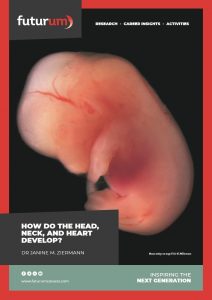
How do the head, neck, and heart develop?
Birth defects affect one in every 33 babies born in the United States each year. A developmental biologist at Howard University in the US, Dr Janine M. Ziermann is studying the head, neck, and heart to find out how head and heart birth defects form.
TALK LIKE A DEVELOPMENTAL BIOLOGIST
Cardiac — relating to the heart
Cardiopharyngeal — relating to the heart and the pharyngeal arches
Cephalic — relating to the head
Congenital — present from birth
Cranial — relating to the cranium
Craniofacial — relating to the skull and the face
Cranium — the skull, especially the part surrounding the brain
Gastrulation — a process during week three of human development, where the embryo transforms from a two-dimensional layer into a three-dimensional structure
Mesoderm — the middle of three layers of an embryo which are the source of many tissues and structures
Palate — the roof of the mouth
Pharyngeal arches — pocket like structures in the embryo that will form most of the head and neck structures
Vertebrates — the group of animals that have a backbone or spinal column, including fish, amphibians, reptiles and birds, and mammals
Despite being in different regions of the body, the muscles of both our heart and our head originate from the same cell group – a group called the cardiopharyngeal mesoderm.
“During embryonic development, the expression of specific genes at precise times and locations ensures that almost all the different head and heart muscles differentiate from this common mesoderm,” says Dr Janine M. Ziermann, who is studying this group of cells for her research at Howard University.
Another cell group, called the cranial neural crest cells, also contributes to the development of the head and heart. During embryonic development, both the neural crest cells and the mesodermal cells create multiple other cell types in the body. Neural crest cells give rise to cells of the nervous system and connective tissue cells. They also generate cells called odontoblasts, chondroblasts and osteoblasts, which eventually help create our teeth and form cartilage and bones in the head.
“The mesoderm forms skeletal structures and muscles,” says Janine. It leads to the creation of the notochord (a rod-like structure that coordinates the backbone development), our urinary and genital systems, and even our axial skeleton (the 80 bones within the central core of our bodies). As well as this, the mesoderm gives rise to cells that differentiate into the heart musculature, blood vessels, muscles and some bones of the head!
By studying both the cardiopharyngeal mesoderm and the cranial neural crest cells, Janine has much of the information necessary to investigate which genes are responsible for causing birth defects in the head and the heart.
What are birth defects?
Structures that are present at birth are called congenital. The terms ‘anomalies’ and ‘defects’ are often used interchangeably and describe something that is different from the expected condition or feature. “Both congenital heart defects and craniofacial anomalies are very common,” says Janine.
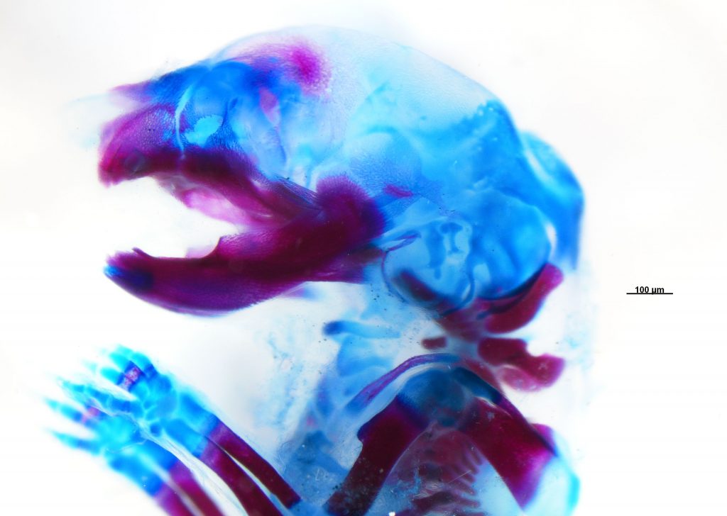
Craniofacial anomalies are deformities which affect the head and facial structure. The most common type of craniofacial anomalies are facial clefts – such as cleft lip or cleft palate – where someone has a separation in their lip or in the roof of their mouth.
Congenital heart defects affect the structure and function of the heart. They can range from mild conditions, such as having a small hole in the heart, to severe conditions, such as poorly formed or missing parts of the heart.
Due to the achievements of modern science, researchers know so much more about the development of these defects now compared to a few decades ago. “However, it is still not clear how even minor changes in the gene regulatory network can have severe effects on the development of head and heart,” Janine explains.
What is Janine’s research?
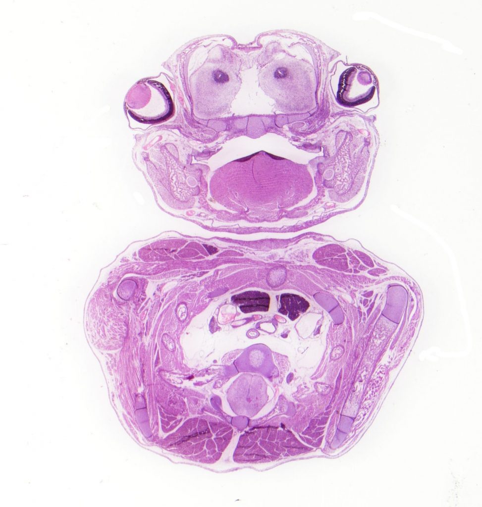
Janine studies tissue interactions needed for the normal development of head and heart musculature, and there are many elements to her research. One of Janine’s research projects focuses specifically on one of the gastrulation-brain-homeobox genes, Gbx2, which is essential for the development of the brain and the spinal cord.
Gbx2 is first expressed during gastrulation. “It is responsible for several important processes and is essential for the movement and survival of neural crest cells,” says Janine. “If Gbx2 is misregulated, the neural crest cells that would usually ensure proper heart development are also likely to be abnormal.”
To carry out this research project, Janine is collaborating with Dr Samuel Waters, a professor at the University of the District of Colombia. Together, they study mouse models that have been genetically modified to have a very low expression of Gbx2.
What have they learnt from the mice?
In order to understand Janine’s results, we first have to know more about the anatomy of the head. On the bottom of our brains, there are 12 nerves called the cranial nerves. The most prominent is the trigeminal nerve, which is responsible for providing sensation to the face and motor function to several muscles. There are three main branches of the trigeminal nerve. One of these is the mandibular branch, which controls the sensations in the jaw, lower lip, gum and, importantly, the motor function of the muscles used for chewing.
“We always learn and teach that the final stage of a muscle to become functional and look normal is the interaction with its nerve,” says Janine. In this case, this means that scientists always thought that the muscles we use for chewing could only be functional and normal once they interacted with the mandibular branch.
When Sam started studying mice with a very low expression of Gbx2, he saw that these mice did not have the mandibular branch of the trigeminal nerve. However, even though the mice did not have a mandibular nerve, they still had seemingly normal looking, but not functional, chewing muscles. This mystery has brought Sam and Janine together as researchers. “This was most fascinating to me!” says Janine. “We are currently looking into the developmental mechanisms that would help us to understand this phenomenon.”
How can mice models teach Janine about human heart defects?
Mice are vertebrates and mammals – just like humans – which means there are many mechanisms and pathways that are very similar, if not the same, between the two. “A mouse heart even looks like a miniature human heart,” says Janine.
Genetic modification techniques allow scientists to manipulate or delete specific genes in mice. “This can tell us the role that the manipulated gene plays in development, which tissues and organs are affected by a misregulation of that gene, and which other genes are changed in their quantity of expression and therefore regulated by the manipulated gene,” explains Janine.
By closely observing the anatomy of a mouse with a specific gene defect, scientists can make educated guesses and investigate potential defects in humans who are missing the same gene. “This process can help future family planning by discussing the risk of the same genetic abnormality happening in the next generation,” Janine adds.
Reference
https://doi.org/10.33424/FUTURUM390
All © JMZiermann
Where might this research go next?
Janine is interested in studying another cell group called the endoderm. “The endoderm sends signals to the developing head and heart,” says Janine. If successful in getting funding for this new project, she will be able to start studying the effect that endodermal cells have on the development of our muscles.
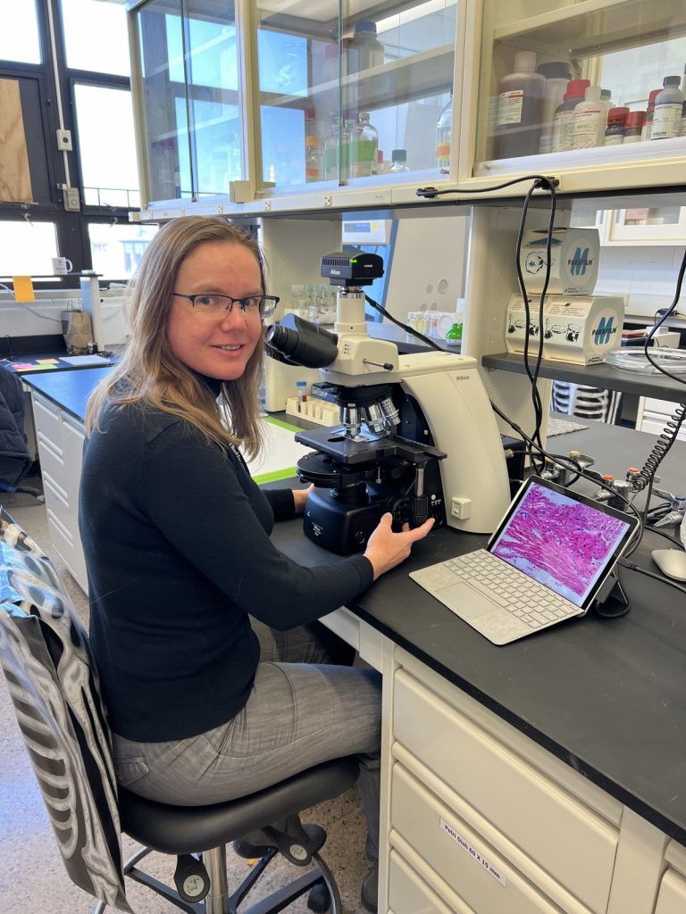
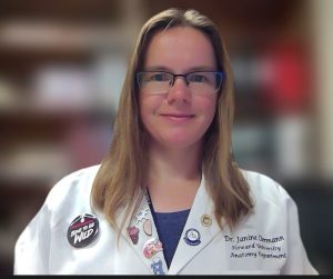 Dr Janine M. Ziermann
HeadHeartEvoDevo-Lab, Department of Anatomy, College of Medicine, Howard University, Washington DC, USA
Fields of research: Anatomy, Developmental Biology
Research project: Understanding cephalic and cardiac development in vertebrates
Funders: Currently, US National Science Foundation (NSF) (previously, Howard University Bridge funding) This work is/was supported by the NSF, under award number #2000005. The contents are solely the responsibility of the authors and do not necessarily represent the official views of NSF.
About developmental biology
Have you ever wondered how all the organs in your body developed and how they all manage to work together? Have you thought about the shapes of your limbs and how they ended up in the positions they are in? These are the areas that developmental biologists explore.
Developmental biologists investigate how animals or plants develop from a single cell to a complex group of cells that all perform separate tasks. They could be studying any process that occurs over the lifetime of an animal or plant, from the very beginning of the reproductive cells, to how the embryo develops and how the body is formed, what the effect of age is, or even what happens within the organism during or after death. To be able to investigate all these processes, developmental biologists must be highly interdisciplinary thinkers who have a good understanding of genetics, biophysics, biochemistry, cell biology, physiology and anatomy.
Scientific advances in this field can have massive impacts on people’s lives. Birth defects, for example, are the reason for one in every five deaths of children who are less than 12 months old. Birth defects can be caused by random chance, inherited gene mutations, complications during pregnancy or birth, or even toxic substances (e.g., fumes, alcohol and medication) that a mother and, hence, the foetus have come into contact with. Developmental biologists, who work to understand how and why these defects happen, distribute the knowledge to health care professionals, who advise families on inherited risks and contribute to developing public policy that allows us to live in healthy, toxin-free environments.
Developmental biology is a very exciting and dynamic field. “The next generation of researchers will have the chance to make organs in a petri dish,” says Janine. Amazingly, this is already being done with cartilage cells and with beating muscle cells! “Perhaps, in the future, we might be able to raise a new heart out of the body cells of the same person, and therefore eradicate organ transplant rejection reactions,” says Janine. “This is still in the future, but not as far as some might think.”
Pathway from school to developmental biology
• During high school and post-16 studies, study STEM subjects such as biology, chemistry, mathematics and physics.
• To work in this field, you should complete a biology-related undergraduate degree such as anatomy, biology, biochemistry, microbiology, or molecular and cellular biology. If you are aiming to work in human anatomy, Janine recommends studying an anatomy degree. However, Janine and many of her colleagues studied biology and ended up working in anatomy.
• Janine recommends taking statistics or comparative anatomy courses during your degree if they are available.
• If you live near Washington DC, see if you can visit Janine’s university. “The Department of Anatomy at Howard University regularly welcomes high school students to visit our anatomy lab and to see first-hand some real human anatomy,” says Janine. “These are usually two-hour sessions with several stations (where students can learn about the skull and other bones, brain, heart, lungs, muscles of the body) and a Q&A session.”
Explore careers in developmental biology
• Developmental biology and anatomy are diverse fields that have a wide range of career opportunities. You might find yourself working in academia or industry as a teacher or researcher or a combination of both, often alongside other scientists and students
from different fields.
• Janine recommends visiting the American Association for Anatomy’s website to read about the latest news within the fields of anatomy and developmental biology. It also provides an introduction to anatomy which explains more about what the field covers.
• Best Accredited Colleges has some helpful information on what a career in developmental biology involves and what the job opportunities are.
• According to Salary Expert, the average annual salary for a developmental biologist in the US is $72,000.
Meet Janine
Who or what inspired you to become a scientist?
I first wanted to be a veterinary doctor. After visiting an introduction session to the veterinary course in Leipzig, Germany, I realised that it might not be for me. I went home and looked for another option. Biology was it.
I wanted to specialise in zoology, and I did, but not in the way I thought in the beginning. Halfway through my education, Dr Lennart Olsson joined our university. He is a great scientist studying the evolution and development of head structures. I did my research rotation with him and fell in love with the subject. I ended up doing both my diploma and PhD in his lab.
As a scientist, it is possible to meet people from all over the world. As long as one keeps an open mind, this brings lots of learning experiences and new friendships. It also provides the opportunity to see different approaches to the same problem.
What is the worst piece of advice you have been given, and how did you ‘unlearn’ it?
‘Figure it out yourself’. This is utter nonsense! I never wanted to bother my professors and lost lots of opportunities because of it. I still think it is not advisable to bother professors with small talk after each lecture; however, if you are interested in a topic and want to learn more, talk to them. They are all human and most actually enjoy talking with students about their lectures and research. If you have a goal and don’t know how to reach it, ask a professor who you trust if they can help outline the next steps. Some say no, and that’s okay, but more will say yes!
Last but not least, it is always okay to say, “I don’t know”. This will be important for your whole career and life. No one knows everything and it is much more respectable to say “Let me look into it” or “Let’s figure that out together” rather than making stuff up.
What are your proudest career achievements, so far?
I got a poster presentation award for my diploma (thesis) and a publication award in 2014 for a paper in my second postdoctoral position. Most recently, I was awarded a US National Science Foundation grant that is supporting the research related to Gbx2. My favourite achievement was that I got to publish two manuscripts with my academic ‘hero’, Dr Drew Noden. Not only did we publish together, but we are still in touch. I was able to visit him before his retirement, and he recently gave a talk at Howard University. I took advantage of him visiting and had long and fascinating conversations with him.
What are your ambitions for the future?
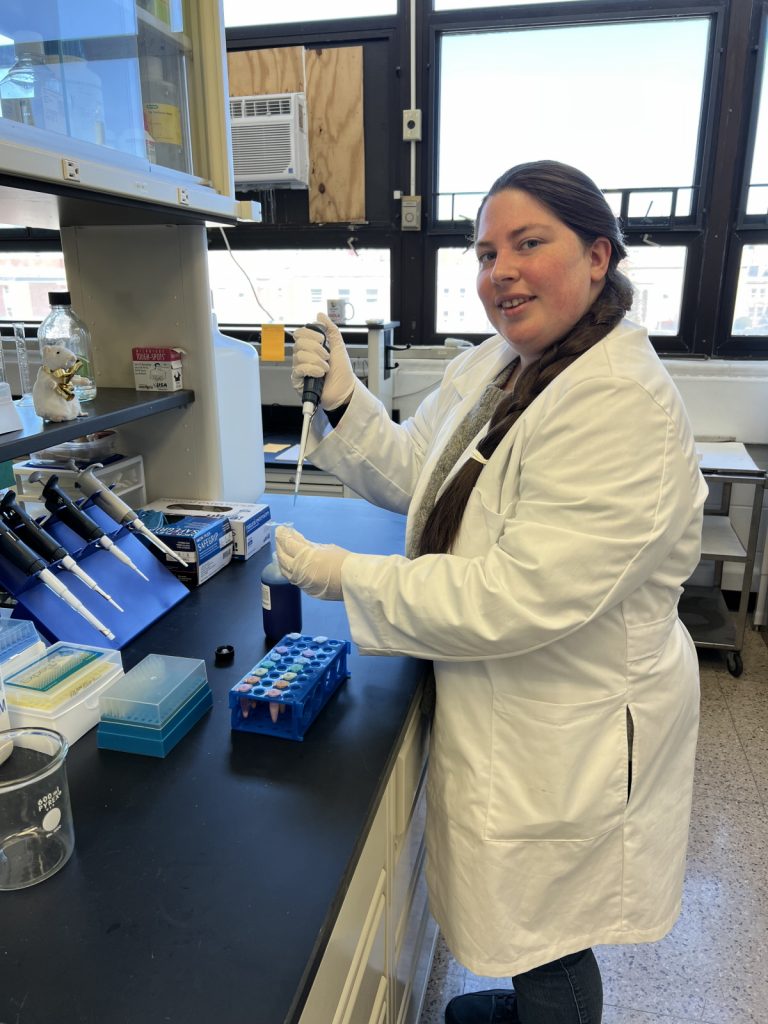
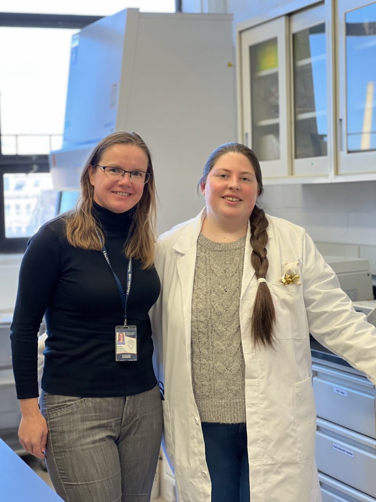
I’m currently trying to stabilise my lab. I recently hired a research assistant/lab manager, Ms Paola Correa-Alfonzo. Getting more funding to keep her and hire MS/PhD students and postdoctoral researchers is next on my plan. With a larger group, I hope to get faster results relevant to the tissue interaction during head and heart muscle development. I also want to establish more collaborations where several people research different aspects on the same model organism. That would reduce not only the number of animals needed in research, but also put changes in gene expression in a larger, more global picture.
Janine’s top tips
1. Find something you are really passionate about, and don’t be afraid to change fields for it!
2. Be flexible. Adding new technologies, new questions and new aspects has its risks but makes things more exciting, not only for yourself but also for funding agencies and the future research and job market.
Do you have a question for Janine?
Write it in the comments box below and Janine will get back to you. (Remember, researchers are very busy people, so you may have to wait a few days.)

