
Making the invisible visible: how can positron emission tomography shine a light on the early stages of disease?
Conditions such as cardiovascular disease, Alzheimer’s disease and Parkinson’s disease can have devastating impacts on the lives of patients and their families. Unfortunately, they are often not diagnosed until far too late. That is why Dr Kiran K. Solingapuram Sai at Wake Forest University School of Medicine in the US is using positron emission tomography (PET) to improve early detection rates, support the development of new treatments and change patients’ lives.
Talk like a medical imager
Alzheimer’s disease – a progressive brain disorder causing memory loss, cognitive decline and changes in behaviour as brain cells become damaged and die
Cardiometabolic disease – a group of interconnected conditions that significantly heighten the risk of heart attacks and strokes
Cardiovascular imaging – a variety of non-invasive and minimally invasive tests, such as PET, used to capture detailed pictures of the heart and blood vessels
Diabetes – a long-term condition where the body cannot properly regulate sugar levels
Microtubules – microscopic protein structures within cells that provide support, help maintain cell shape and transport materials throughout the cell
Neurodegeneration – the gradual loss of structure or function of nerve cells in the brain and nervous system
Parkinson’s disease – a progressive neurological disorder caused by the loss of dopamine-producing brain cells
Positron emission tomography (PET) – a medical imaging technique that uses radiotracers to visualise biological activity within the body
Radiotracers – molecules labelled with a radioactive substance that is detectable by a PET scanner, allowing scientists and doctors to study specific biological processes in the body
Medical imaging techniques such as X-ray and magnetic resonance imaging (MRI) have revolutionised medicine, allowing us to see inside the body and identify problems such as fractures, tumours and organ abnormalities. Positron emission tomography (PET) goes one step further, enabling us to visualise the biological activity of diseases before they cause any visible structural change.
“Disease often changes how the body functions long before it changes how it looks,” says Dr Kiran K. Solingapuram Sai from Wake Forest University School of Medicine. “PET is a powerful technique that allows us to see what is happening inside the body at a molecular level.” PET is now a standard clinical tool used in oncology (the study of cancer), neurology, cardiology and the prevention of cardiometabolic disorders. For example, in cardiology, PET can measure blood flow to the heart muscle, detect inflammation, and assess viability of heart tissue after a heart attack.
The process is relatively simple. “A patient is injected with a tiny amount of a radioactive molecule, called a radiotracer, that travels through the bloodstream to specific areas of interest,” explains Kiran. “A PET scanner captures signals from the radiotracer and converts them into three-dimensional images, allowing us to determine where the radiotracer has accumulated in the body. The result is a detailed map of biological activity.” In other words, PET lets us see biology in action.
How are radiotracers developed?
“Radiotracers are specially designed molecules that target a specific biological process in the body,” says Kiran. “They are like ‘tracking devices’ that bind to a particular protein, receptor or metabolic pathway.”
The observation of different biological activities requires different radiotracers as a PET scan can only show the specific biological process or molecule that its radiotracer is designed to detect. For example, a radiotracer designed to measure how cells use glucose cannot be used to reveal cancer-related proteins. “Developing new radiotracers is critical because the targets we want to image keep changing,” says Kiran. “Each new radiotracer unlocks a new door.”
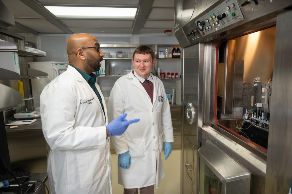
Kiran creates radiotracers using a long, iterative process, bringing together specialists in bioinformatics, chemistry and biology. Once a biological target is chosen, such as a protein linked to a specific disease, Kiran designs molecules that bind to it and chooses the most effective one. “After choosing molecules, we attach a radioactive label to them,” says Kiran. “We then test them in cells and animal models to confirm they are reaching the target and staying there long enough to image. If the results are promising, we move towards testing them in monkeys followed by human trials.”
PET and neurodegenerative disease
PET plays a vital role in the detection and treatment of neurological disorders, such as Alzheimer’s disease, Parkinson’s disease and stroke. “These diseases begin at the molecular level long before symptoms appear,” says Kiran. “If we can design tracers that detect these early changes, we can diagnose diseases earlier and track treatment responses more accurately.”
“Neurodegeneration is a slow, devastating process, and for too long we have been diagnosing diseases only after significant damage has already occurred,” continues Kiran. “But now we can image important proteins of Alzheimer’s disease, detect dopamine deficits in Parkinson’s disease and assess brain metabolism after stroke or traumatic injury.”
An early feature of both Alzheimer’s disease and Parkinson’s disease is the degradation of microtubules – hollow structures that maintain the shape of cells, transport nutrients and support communication between cells. “Our lab developed the first PET radiotracer that can enter the brain and track microtubule integrity in living animals and humans,” says Kiran. “We are investigating additional applications for it in Alzheimer’s patients.”
Kiran and his team have also developed a PET radiotracer for a receptor that is disrupted during Alzheimer’s disease. “Being able to image this receptor gives us an entirely new way to understand neurological disorders and allows us to test whether new drugs that target this receptor are working,” says Kiran.
PET and cardiometabolic diseases
Kiran is also developing new ways for PET to contribute to the treatment of chronic cardiometabolic diseases such as diabetes, which can lead to heart disease. “Type 2 diabetes damages blood vessels and the heart over time, which can lead to heart failure in diabetic patients,” says Kiran. “With PET, we can detect inflammatory processes developing in blood vessels and organs like the lungs and joints, often before conventional tests would raise any alarm.”
Kiran has developed a new radiotracer for a receptor that is found in the pancreas, gut and liver and is important in the regulation of blood glucose and insulin release, called GPR119. “Pharmaceutical companies have been actively trying to develop drugs that reach and activate this receptor to treat diabetes,” says Kiran. “Our radiotracer allows us to directly evaluate how the activity of this receptor changes with diabetic conditions and, more importantly, how well potential drugs engage with it.”
Reference
https://doi.org/10.33424/FUTURUM721
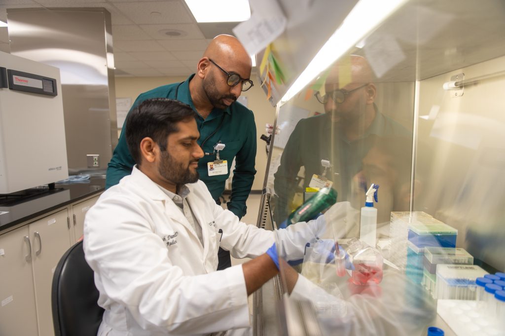
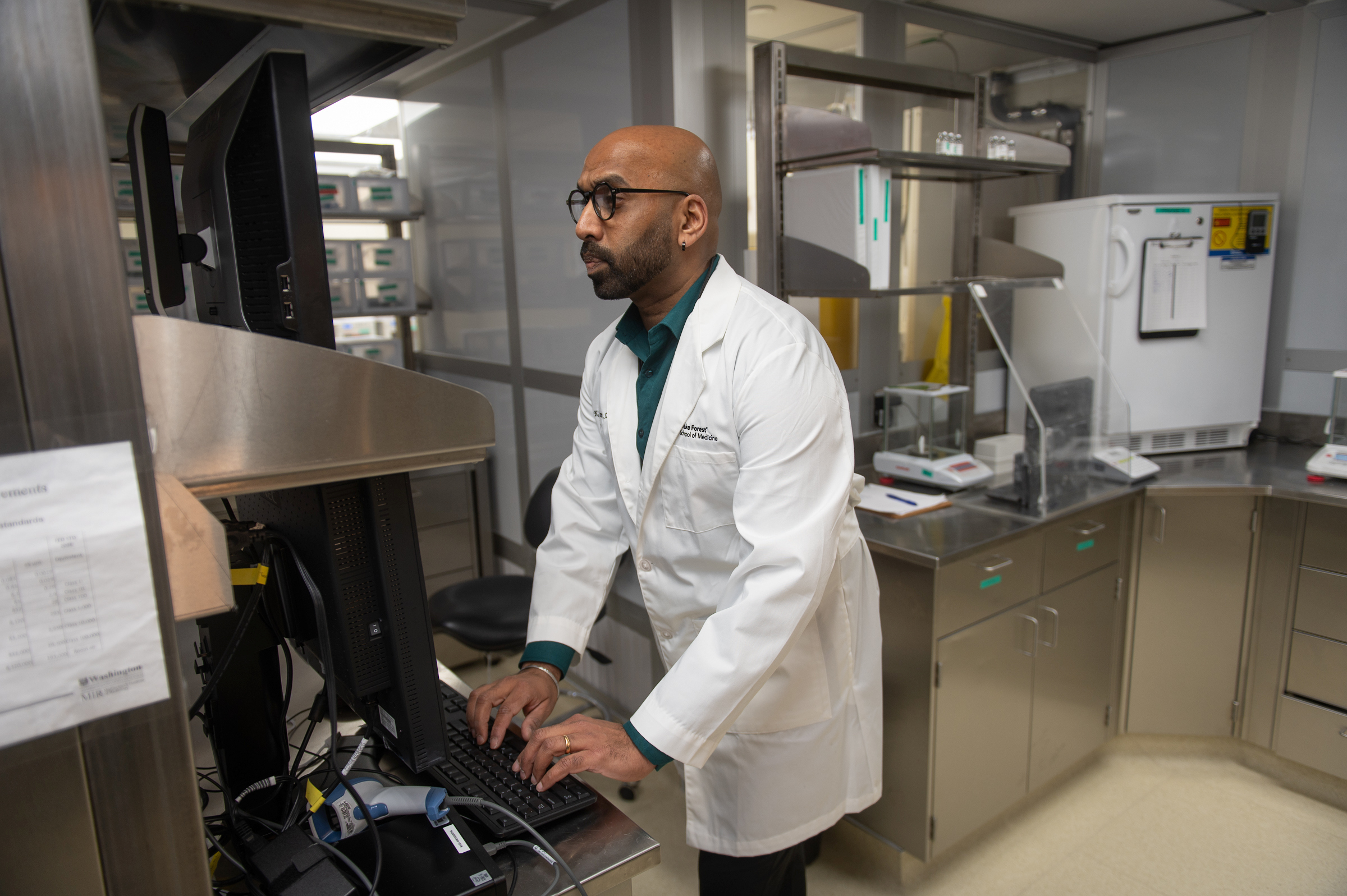
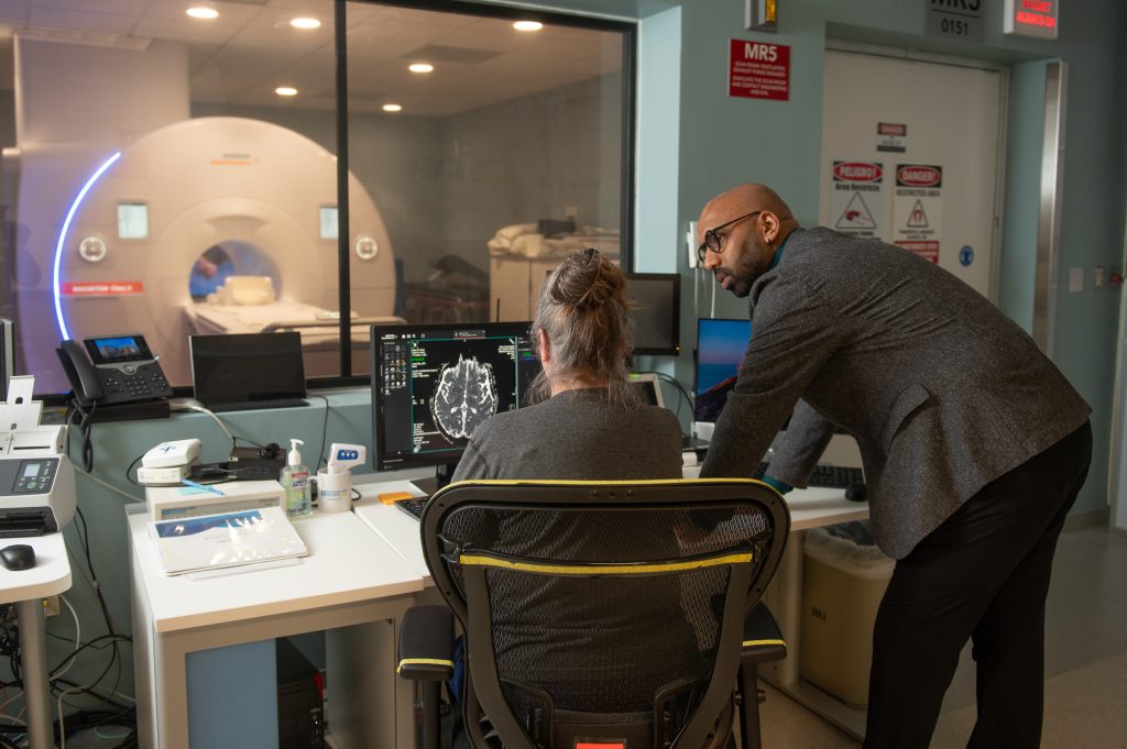
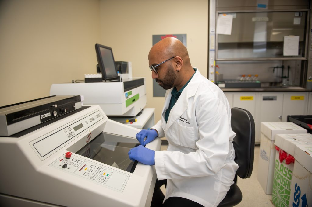
Kiran working on autoradiography with post-mortem brain tissue analyses.


This educational material has been produced by Wake Forest University School of Medicine in partnership with Futurum and with grant support from The Duke Endowment. The Duke Endowment is a private foundation that strengthens communities in North Carolina and South Carolina by nurturing children, promoting health, educating minds and enriching spirits.
Let us know what you think of this educational and career resource. To provide input, simply scan the QR code or click here.
Imaging impact
Ultimately, Kiran’s goal is to improve patients’ lives by finding new ways to detect diseases at earlier stages and by helping doctors choose treatments that are tailored to each individual. “If we can detect Alzheimer’s disease five or ten years before symptoms appear, that changes everything for patients and families,” explains Kiran. The same principle applies to diseases such as Parkinson’s, diabetes and cancer, where earlier detection can lead to better outcomes. “The work we are doing is opening up entirely new biological windows,” continues Kiran. “We are developing new radiotracers for neurological diseases, such as Alzheimer’s disease and alcohol use disorder, and for cardiovascular diseases. We are developing new molecular imaging tools to make the invisible visible.”
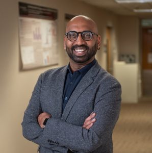 Dr Kiran K. Solingapuram Sai
Professor, Department of Radiology, Wake Forest University School of Medicine, USA
Field of research: Medical imaging
Research project: Imaging new markers for early detection of Alzheimer’s disease
Funder: US National Institutes of Health (NIH)
About medical imaging
Medical imaging allows us to see inside the body, helping doctors diagnose disease, monitor treatment and understand how our bodies work. As an area of medicine, it offers a huge range of opportunities, from developing new radiotracers and treatments to improving the sensitivity of scanners and building innovative AI tools to support the interpretation of images.
“What makes this field exciting is that it sits at the intersection of chemistry, physics, biology, medicine, engineering and technology,” says Kiran. “No matter where your strengths lie, whether you love working with patients, writing code, managing teams or discovering new science, there is a meaningful place for you here.”
The main challenge for Kiran is the length of time it takes to get new radiotracers from the laboratory to the patients. “You can develop a beautiful molecule that works perfectly in rodents and then spend years navigating the regulatory and manufacturing hurdles required to use it in humans,” explains Kiran. “Science often moves faster than the systems built to verify and translate it. That tension between scientific possibility and practical translation is the challenge I grapple with most.”
However, the reward of seeing a new radiotracer work for the first time makes it all worthwhile. “After months or years of chemistry and preclinical testing, that moment is electrifying,” says Kiran. “There is nothing quite like seeing science work. But the deepest reward is knowing that this work is helping patients. When I think about the patients and families affected by chronic disorders such as cardiometabolic diseases and Alzheimer’s disease, I think about what earlier, more accurate diagnosis could mean for them. That is what keeps pushing me every day – I am contributing to making the world a better place!”
Pathway from school to medical imaging
Studying chemistry, biology and physics at school and beyond will build a valuable foundation. “Never lose sight of biology,” says Kiran. “Understanding how the body works and how disease begins at the cellular level – that’s what gives the science its purpose. The best people I’ve worked with are the ones who can think like a chemist and a biologist at the same time. You need both.”
“At university, chemistry, biochemistry, pharmacology, biomedical engineering and physics are all great pathways into the field,” says Kiran. “I’d also strongly encourage students to explore computer science or data science. AI is changing how we read and interpret imaging data, and that’s only going to grow.”
“Medical imaging is not a single discipline; it’s chemistry, physics, biology and data all talking to each other,” says Kiran. “The more ‘languages’ you speak, the further you’ll go.”
Explore careers in medical imaging
“My first advice is simple: find a lab and get involved,” says Kiran. “Reading about science is one thing – doing it is something else entirely. The moment you pipette your first compound, run your first scan or see a PET image light up a region of the brain, something clicks. That experience is irreplaceable.”
“In my lab at Wake Forest, we actively welcome motivated high school students and undergraduates for summer volunteering,” says Kiran. “Students get to work alongside our team on real neuroscience. Some students who began with us as summer volunteers have gone on to medical schools and graduate programmes at some of the country’s finest institutions.”
“The Society of Nuclear Medicine and Molecular Imaging and the World Molecular Imaging Society are the leading professional organisations in our field,” says Kiran. “They offer educational resources and host annual meetings bringing together researchers, clinicians, and students worldwide.”
Meet Kiran
I grew up in India in a lower middle-class family. Every step of the way, I depended on the generosity of people around me — family, friends, neighbours, teachers, community programmes and strangers who believed in a kid they barely knew. It taught me that where you start does not determine where you finish, and that nobody gets anywhere alone.
I lost my grandmother to dementia when she was only 64. It came on fast and it was devastating – we lost her within two months of her diagnosis. Through all of it, there were no tools to catch it early, no way to see what was happening inside her brain and, where we lived in India, no real access to specialised care even if those tools had existed. That experience never left me. Every PET tracer we develop, every image we generate, every earlier detection we inch toward – it is for her. It is for every family sitting where my family sat, watching someone they love slip away while medicine looks on without the tools to intervene.
Every radiotracer we develop starts with a biological question and then becomes an enormous, multi-layered puzzle. It takes collaboration across chemistry, biology, physics and clinical medicine to develop them. Some of the best ideas in my lab have come from conversations between a chemist and a clinician who are both looking at the same image and asking completely different questions. That collision of perspectives is where the science gets exciting.
I genuinely believe that curiosity about the world makes you a better scientist. When you sit with people whose lives look nothing like yours, you come back to the lab with a broader sense of what matters and who you are ultimately doing this work for.
Spending time with my family is what truly recharges me. My beautiful wife, Mohini, and my two cute daughters, Kiara and Kiona, have a way of pulling me completely into the present moment – and that is something no amount of data or grant writing can replicate.
Dance genuinely lights me up. I have been learning Bharatanatyam, one of the oldest and most beautiful classical dance forms from India, and it has become something I deeply treasure – the discipline, the storytelling, the way every movement carries meaning. In many ways, it reminds me of science. Both require patience, precision and a willingness to keep practising long before mastery arrives.
Kiran’s top tips
1. Get comfortable with failure. The scientists who make the biggest contributions are the ones who have the resilience to keep going when things don’t work, and the wisdom to learn something useful from every failure.
2. Find mentors who believe in you and push you. I have been fortunate throughout my career to work with scientists who challenged me to think bigger and to not be satisfied with easy answers.
3. Remember why you started. The reason for pursuing this kind of science is the impact it can have on real people’s lives. On the days when experiments fail, holding on to that purpose makes all the difference.
Do you have a question for Kiran?
Write it in the comments box below and he will get back to you. (Remember, researchers are very busy people, so you may have to wait a few days.)

Learn about how we medical imaging can advance treatment:
0 Comments