Creating a clear image of myopia: discovering the causes and developing treatments
Myopia, also known as near-sightedness, causes blurry distance vision and is estimated to affect over 30% of the world’s population. At Emory University School of Medicine in the US, Professor Machelle Pardue and her research team are working to better understand the underlying causes of myopia. They hope their discoveries will identify novel therapeutic interventions for treating myopia in children.
Talk like an ophthalmologist
Axial length — the length of the eye, usually measured from the front of the eye to the back of the retina
Emmetropisation — a process that occurs after birth whereby the refractive components of the eye and the axial length come into balance to achieve clear vision
Myopia (near-sightedness) — a type of refractive error that typically occurs when the eye is too long and light focuses in front of the retina, causing distant objects to appear blurry
Ophthalmology — the branch of medicine focused on the eye and vision
Optical power — the degree to which a lens or other optical system bends or focuses light
Photoreceptors — the cells in the retina that are responsible for detecting light
Refractive error — a common type of vision problem that occurs when light does not focus correctly on the retina, resulting in blurry vision
Retina — the light-sensitive layer of tissue in the back of the eye
Sclera — the white, outermost layer of the eye that provides structural support
Myopia is characterised by blurry distance vision. It is a very common eye condition that currently affects over 30% of the world’s population, and its prevalence is rapidly increasing. It is predicted that by the year 2050, 50% of the world’s population (equivalent to almost 5 billion people) will have myopia. Glasses and contact lenses can correct the visual symptoms of myopia by helping people to see clearly, but they do not stop the progression of the condition or prevent the associated vision-threatening complications that can lead to visual impairment.
How does myopia occur?
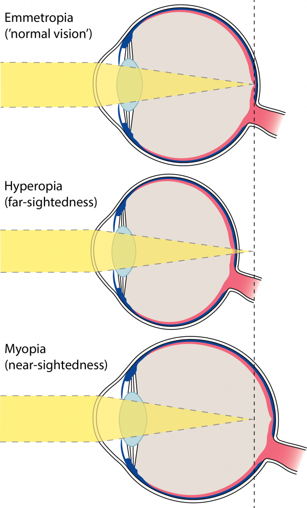
After birth, the optical power (i.e., focusing strength) and size of our eyes come into balance to achieve clear vision, a developmental process known as emmetropisation. However, if the eye’s optical power and axial length fail to come into balance, then light does not focus correctly on the retina, the light-sensitive tissue in the back of the eye. This leads to refractive errors, which result in blurry vision. The two main types of refractive error are myopia (near-sightedness), which typically occurs when the eye is too long, and hyperopia (far-sightedness), which typically occurs when the eye is too short.
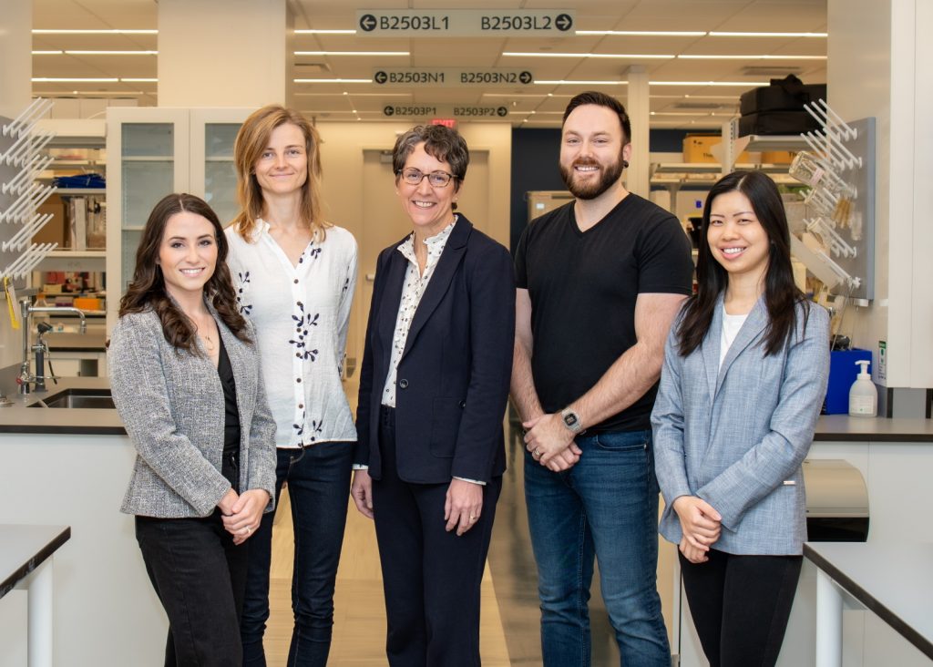
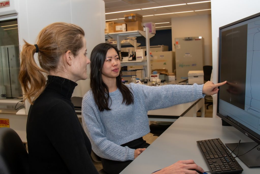
At Emory University School of Medicine, vision scientist Professor Machelle Pardue is working with her research team: Dr Reece Mazade, Dr Melissa Bentley-Ford, Dr Linjiang Lou and Dr Teele Palumaa. They are improving our understanding of the mechanisms that cause myopia to help reveal novel therapeutic interventions for the treatment of this condition in children.
What influences eye growth and myopia development?
It is well established that both genetic and environmental factors are involved in eye growth and myopia development, although the exact mechanisms remain unknown. Scientists have found many genes associated with myopia, and it is known that children with parents who are myopic (near-sighted) are more likely to develop myopia. The major environmental factors associated with myopia are years spent in education and time spent outdoors. More education is associated with more myopia, whereas spending more time outdoors protects against myopia development.
Light exposure plays a critical role in eye growth and myopia development. The lighting environment we are exposed to indoors is very different from the natural light we typically experience outdoors. “The intensity of light is important,” explains Machelle. “We have found that bright light and dim light are both important for protecting against myopia development, and that myopic children spend less time in both of these lighting conditions.”
Light is also important for regulating circadian rhythms, which are internally driven changes within our body that oscillate with an approximate 24-hour cycle. “Keeping a regular light/dark schedule is highly important for maintaining healthy circadian rhythms,” explains Teele. “In fact, several lines of evidence suggest that circadian rhythms are associated with myopia development, and I am studying the mechanisms behind this association.”
What is the team investigating?
Visual signals from the environment are processed within the eye to control eye growth and guide emmetropisation. These visual signals are detected and processed by the retina, initiating a chain of events that are then detected by the sclera, the white outermost layer of the eye, to drive changes in eye size. Machelle’s lab is studying the retinal pathways and the roles of two signalling molecules, retinal dopamine and retinoic acid, in the regulation of eye growth and myopia development.
Retinal dopamine is critical for many aspects of light-dependent functions in the retina, and it has also been shown to be involved in myopia development. “Retinal dopamine levels are reduced in most animal models of myopia, and we have shown that increasing retinal dopamine levels can prevent experimental myopia in animals,” Machelle explains.
Retinoic acid is highly important for regulating many developmental and functional processes within the eye and other organs. In contrast to retinal dopamine, retinoic acid is increased after experimental myopia, and increasing retinoic acid levels can cause myopia in animal models.
How does the team study the mechanisms behind myopia development?
Machelle has established a mouse model of myopia to study the retinal and scleral mechanisms involved in myopia development. “An advantage of the mouse model is that we can manipulate both the genetics and environment,” Teele explains. “We can study the impact of a specific gene on myopia development by inactivating or removing it from the genome of the mouse.”
Reference
https://doi.org/10.33424/FUTURUM452
When axial (eye) length is correct (top diagram), light focuses on the retina (red layer) causing normal vision. If the eye is too short (middle diagram), light focuses behind the retina causing hyperopia, whereas if the eye is too long (bottom diagram), light focuses in front of the retina causing myopia
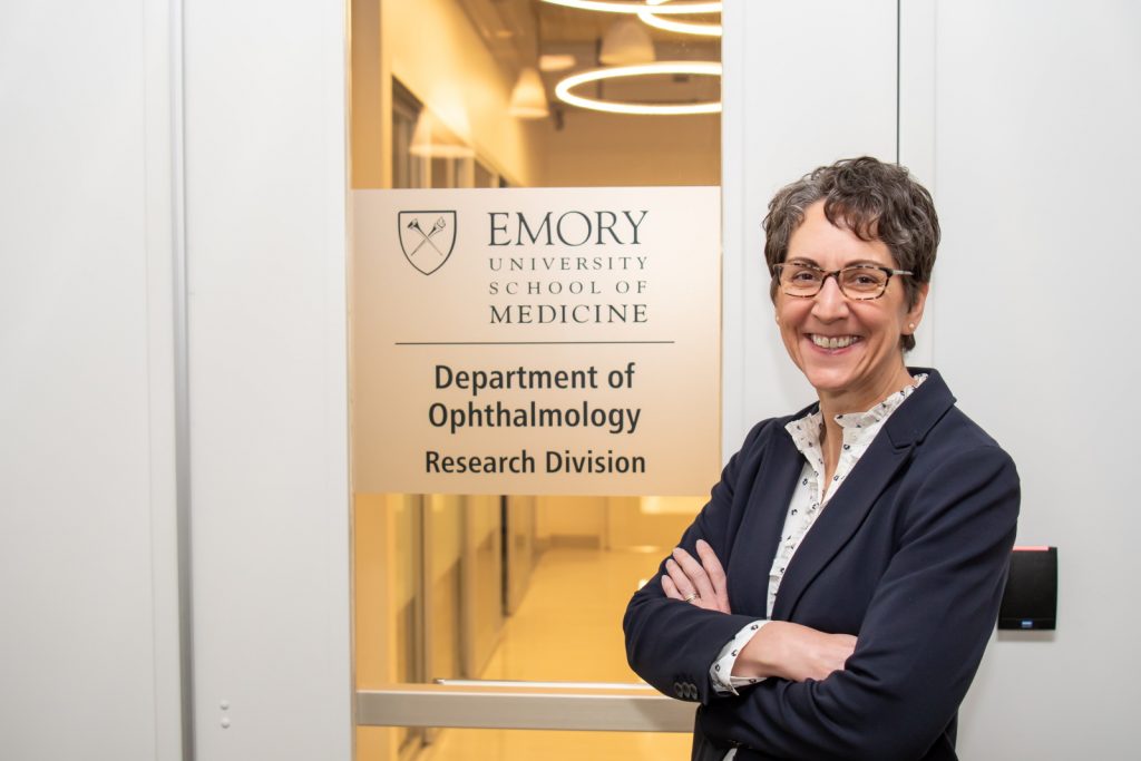
Machelle is a Professor of Ophthalmology at Emory University School of Medicine
All images © Pardue Lab
Experimental myopia can be induced in animal models by placing a negative powered lens or a translucent light diffuser in front of the eye, both of which cause the eye to grow longer. “We use custom instruments to measure the refractive error and axial length of the mice, which allow us to examine how normal eye growth or the response to experimental myopia is affected with different genetic or environmental manipulations,” explains Linjiang. “We also measure whether retinal dopamine levels are altered by these experimental conditions.”
“Previous work in the lab has shown that rod photoreceptors, which are responsible for vision in dim conditions (such as at night), play a critical role in refractive development, whereas cone photoreceptors, which function under brighter daytime conditions and are responsible for sharp vision, are less critical,” Reece says. “I perform electrical recordings to measure the response of individual cells in the retina to further examine which retinal pathways are affected in experimental myopia.”
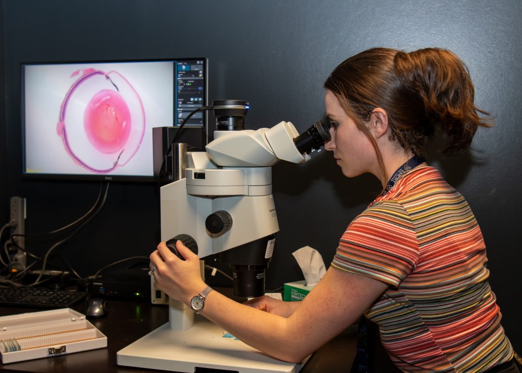
More recent work from Machelle’s lab has demonstrated that the stiffness of the sclera is decreased in experimental myopia and with retinoic acid treatment. “Ultimately, changes in eye size are due to remodelling of the sclera,” explains Melissa. “I use high-resolution microscopes to visualise cells in the sclera and examine how they are affected during experimental myopia. This will allow us to better understand the role of these cells in myopia.”
How can Machelle’s research be applied to therapeutic interventions for children?
Current treatments for myopia can slow its progression, but we do not know how these treatments work. “Identifying the underlying mechanisms responsible for eye growth and myopia development helps us to identify potential targets to develop better treatment interventions for this condition,” says Machelle. “For example, light therapies that stimulate retinal pathways could be a practical and non-invasive intervention to target myopia progression in children.” This is already an emerging topic of interest in the field, and research findings from Machelle’s lab will help inform the development of these therapeutic interventions for treating myopia in children.
 Professor Machelle Pardue
Vice Chair and Director of Research, Department of Ophthalmology, Emory University School of Medicine, USA
Field of research: Ophthalmology
Research project: Investigating the retinal mechanisms of eye growth and myopia development
Funder: US National Institutes of Health (NIH)
About ophthalmology
Ophthalmology is the study of eyes and vision. It includes the study of the structure and function of the eye, how visual function is affected by eye diseases and disorders, and how to prevent and treat eye conditions. “Ophthalmology is a multidisciplinary field, and interdisciplinary collaboration is essential for solving complicated problems. It allows us to look at problems from various knowledge bases and perspectives,” says Machelle. “For instance, I am an expert in retinal function and anatomy, and I collaborate with experts in cell biology and development, ocular biomechanics, retinal circuitry and other fields to advance our research in innovative ways.”
Opportunities and challenges in ophthalmology research
One of the most rewarding aspects of research is the opportunity to work with a team of intelligent and talented individuals. Each person in a research group provides their unique set of talents and expertise. “It is important to pursue ideas and concepts that interest you. You never know how your expertise can be applied to a new problem or idea,” says Melissa. “There are opportunities for basic science research to study the mechanisms underlying eye diseases and to develop treatments for these diseases. Clinical research then applies the knowledge we learn to improve patient care.”
“The eye is a wonderous organ!” says Machelle. “Its neurons, muscle, lens and collagen components are all easily accessible through the front of the eye. I find it fascinating to study the perfect coordination of these different components that allow us to see our world. However, it can be challenging to study such a complex organ. There are multiple aspects of the eye and eye health that we are still learning about.”
Pathway from school to ophthalmology
• At high school and beyond, study biology, physics, chemistry and mathematics.
• Completing a bachelor’s degree in a biomedical science field, such as neuroscience, physiology, biology or anatomy, provides a good foundation to help understand basic concepts that can be applied to ophthalmology and vision research.
• “The best way to gauge your interest in ophthalmology and vision research is to do a research project in a laboratory that conducts vision research,” says Linjiang. “You will have the opportunity to get hands-on experience in a laboratory setting and learn about vision science.” Many colleges and universities have courses that allow you to conduct research in a laboratory for course credit.
• Machelle recommends exploring Webvision (www.webvision.med.utah.edu) to learn some basics about the eye. “The National Eye Institute (NEI, www.nei.nih.gov) and Association for Research in Vision and Ophthalmology (ARVO, www.arvo.org) websites are also useful. ARVO has events for members-in-training, including for students, postgraduate fellows and residents,” Machelle advises.
Explore careers in ophthalmology
• To become a vision scientist, you should complete a graduate degree, then undertake post-graduate training in a vision research laboratory. You will work in a laboratory to conduct research focused on improving our understanding of vision and eye conditions. This could be at an academic institution or in industry at a private research company.
• To become a clinician specialising in eyes and vision, you will need to complete a medical degree and residency to become an ophthalmologist, or an optometry degree to become an optometrist. Then, you can also do additional training or fellowships to specialise in different areas of the field.
Meet Machelle’s research team:

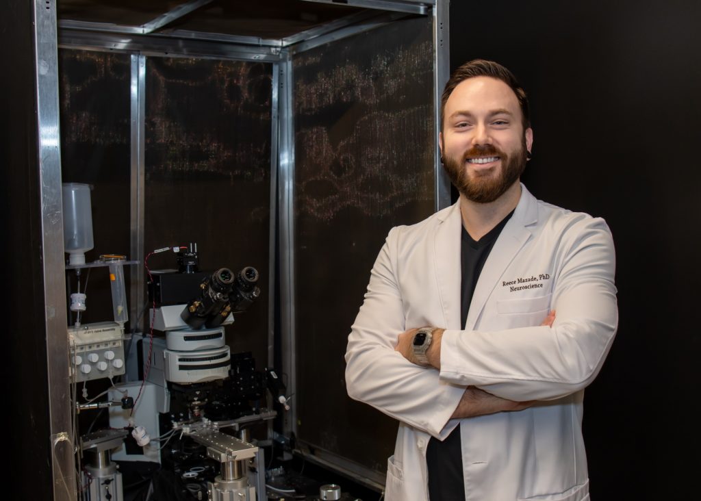
Dr Reece Mazade
Research Scientist
I’ve always been curious about nature, especially animal biology. In high school, an amazing biology teacher encouraged me to submit a scientific research project on bird ecology to the state university, and this experience jump-started my scientific career.
After learning about physiological systems during my biological sciences degree, I became interested in sensory neuroscience. I pursued a PhD in physiological science, investigating how retinal neurons signal in different lighting conditions. As I started applying my findings to vision and eye disorders, my studies led me to the field of ophthalmology.
I love discovering how our eyes translate visual input from the natural world into something we can perceive. The eye’s remarkably organised structure and complexity make it an exciting organ to study!

Dr Melissa Bentley-Ford
Postdoctoral Fellow
At an early age, I decided that I not only wanted to learn about the world around me but also to discover how it worked. As a scientist, I can do just that.
After obtaining my bachelor’s in biochemistry and PhD in cell and developmental biology, I jumped into the world of ophthalmic research. My background gives me expertise in cell signalling, genetics and molecular biology. By combining this with my colleagues’ expertise in neuroscience, physiology, engineering, optics and clinical research, we can tackle questions from different angles.
I became interested in ophthalmology research because I was allured by the fascinating biological questions and complexities that exist in the eye. Every day, I get to wake up and work to discover mechanisms that allow us to see.

Dr Linjiang Lou
Postdoctoral Fellow
My interest in research started during my undergraduate studies in neuroscience. I had a great professor whose passion and excitement for the topic and for research inspired me to pursue a career as a scientist.
I was introduced to vision science and ophthalmology during my PhD in physiological optics. I find vision research fascinating because the eye is so complex and so interconnected with the brain. My research training has focused on eye growth and myopia, a constantly growing field with many questions yet to be answered.
Ophthalmology is a multidisciplinary field. We interact with scientists with diverse backgrounds and expertise, which allows us to approach questions from different perspectives. Having good mentors along the way has helped formulate my perspectives and experience in the field.

Dr Teele Palumaa
Postdoctoral Fellow
My curiosity about how biological processes function in health and disease, particularly in diseases that are a significant burden to societies, inspired me to become a scientist.
While studying medicine, I realised that I was equally interested in science, especially in how our bodies have circadian rhythms. After graduating from medical school, I earned a PhD in clinical neurosciences, conducting research on the circadian rhythms of the retina. Since then, I have been combining clinical life with scientific research. I started an ophthalmology residency programme and with one final year to go, I took a break from clinical work to do science on a post-doctoral level.
I enjoy ophthalmology research because I can make an impact by studying problems that matter on a global scale.
Do you have a question for the team?
Write it in the comments box below and the team will get back to you. (Remember, researchers are very busy people, so you may have to wait a few days.)
Learn how ophthalmologists are preventing diabetes-related vision loss:
www.futurumcareers.com/how-can-diabetes-lead-to-vision-loss-and-how-can-this-be-prevented


0 Comments