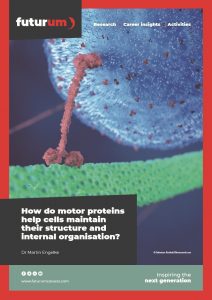
How do motor proteins help cells maintain their structure and internal organisation?
How do cells receive developmental cues and other specialised signals? How do your airways prevent dangerous particles from reaching your lungs? And how do sperm swim? The answer to all these: cilia. In the Cellular Organization Lab at Illinois State University in the US, Dr Martin Engelke and a team of cellular and molecular biologists are investigating the role that motor proteins play in building and maintaining these crucial cell structures.
Talk like a cellular and molecular biologist
Cell signalling — the processes by which a cell exchanges messages with other cells and perceives its environment
Cilia — tiny hair-like protrusions that extend from a cell membrane
Cultured cell — a live cell grown artificially in a petri dish in a laboratory
Intraflagellar transport (IFT) — the process by which motor proteins transport molecules inside cilia
Kinesin-2 — a transporting motor protein involved in various intracellular transport processes, including IFT
Microtubule — a polymeric structure that, as part of the cytoskeleton, supports the shape of a cell and functions as roadways for intracellular transport
Motor protein — a type of protein that can move along the cytoskeleton of cells
Cilia (meaning ‘eyelashes’ in Latin) are tiny hair-like protrusions that extend from a cell membrane, supported by a microtubule structure. Cilia can be divided into two forms: motile and non-motile. Non-motile cilia, which cannot move, are found on nearly every type of animal cell. They play a crucial role in cell signalling, by receiving extracellular signals and passing these ‘messages’ to the cell. Motile cilia exist on specialised cells to fulfil specific functions. On stationary cells, the beating motion of motile cilia moves fluid. For example, cilia on cells lining the respiratory tract move fluid over the surface of the airways, while cilia in the female reproductive tract move egg cells from the ovary to the uterus. The motion of some motile cilia, also known as flagella, can propel non-stationary cells forward, for example on sperm cells.
How are cilia maintained?
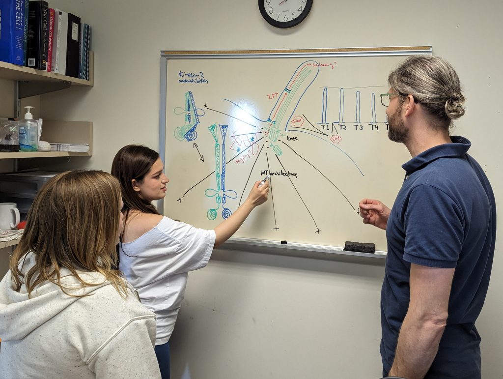
To build and maintain cilia, molecules are transported along the ciliary microtubules by a process known as intraflagellar transport (IFT). Motor proteins power IFT by literally ‘stepping’ along the ciliary microtubules and carrying ciliary building blocks and signalling molecules with them. “IFT motor proteins move large protein complexes into and out of cilia, with kinesin-2 powering IFT towards the ciliary tip and dynein-2 powering IFT towards the ciliary base,” explains Dr Martin Engelke, a cellular and molecular biologist at Illinois State University. “When viewed under an electron microscope, the protein complexes that kinesin-2 transports resemble an assembly of train wagons, into which protein cargo can be loaded to be imported to or exported from the cilia.”
How is Martin’s team studying kinesin-2?
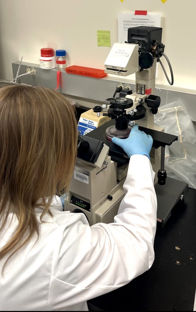
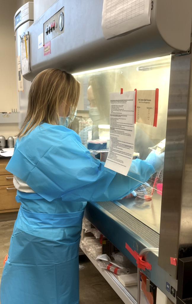
Biologists in Martin’s Cellular Organization Lab are examining the role of kinesin-2 in cilia generation and maintenance. One method involves using gene editing tools to remove the genes that produce kinesin-2 from cultured cells. “These cells do not produce kinesin-2 and, thus, cannot generate cilia,” explains Martin. The researchers then add kinesin-2 genes back into the cells, at which point they regain the ability to make cilia and their regrowth can be observed under a microscope. This method is useful because the researchers can add mutant versions of the kinesin-2 genes, allowing them to observe the function of different versions of kinesin-2, which may cause the cilia to grow longer or shorter, or prevent cilia from growing at all.
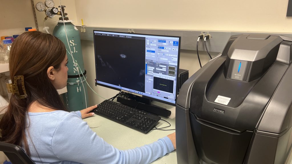
Lindsey Margewich, a PhD student in the lab, allows cultured cells to grow cilia and then inhibits kinesin-2 function by using a drug that binds to the motor protein and prevents it from moving on the microtubules. The cells lose the ability to maintain their cilia when the motor becomes inhibited. “I incorporate fluorescently tagged proteins into ciliary membranes and use a fluorescent dye to stain ciliary microtubules,” she explains. “This allows me to see them under a microscope. I take photos of these live cells every two minutes over the course of a few hours to see how the cilia change after I have inhibited kinesin-2.”
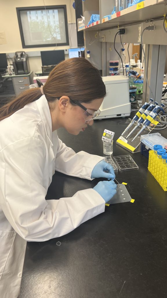
Farimah Mohammadi, another PhD student, hopes to determine how kinesin-2 molecules reach the base of cilia, the starting point of IFT. “Theoretically, the molecule might navigate itself, it might be transported by another motor protein, or it might diffuse through the cell’s cytoplasm to reach the ciliary base,” she explains. To examine these possibilities, Farimah will use a technique called fluorescence recovery after photobleaching. “This involves using a laser to bleach a small region of fluorescently tagged proteins, followed by monitoring the recovery of fluorescence over time as unbleached proteins move into the bleached area,” she says. “By fusing the kinesin-2 motor protein with a fluorescent marker, I will also be able to track its movement kinetics and observe how it behaves within the cellular environment.”
What has the team discovered?
The team’s gene replacement work has indicated that altering the properties of kinesin-2 alters the characteristics of cilia, for example, causing them to grow longer. Also, while it had been previously discovered that cilia disappear when IFT is impaired, it was not known how they vanish. For the first time, Lindsey has observed how cilia disassemble when kinesin-2 is inhibited in live mammalian cells. “The cilia are usually shed from the cell, but sometimes they shrink,” she says. “Within six hours of inhibiting kinesin-2, all cilia that had formed on our cultured cells had disappeared.” The team’s experiments have confirmed that ongoing IFT is needed to maintain the ciliary structure in most cells, but the reasons for this are still unknown.
What next?
Once Farimah has discovered how motor proteins reach the base of cilia, she will shift her focus to investigate what mechanisms are responsible for activating kinesin-2 when it reaches this site. Lindsey is now trying to determine what causes cilia to disassemble when kinesin-2 is no longer driving IFT. “Once we determine how cilia disassemble in the absence of IFT, we can hypothesise what proteins or processes might be responsible and study their role in the disassembly,” she says.
When cilia malfunction, it can lead to a range of diseases, including retina degeneration, kidney disease and cancer. Therefore, it is of great importance to understand how cilia, and the motor proteins which govern them, function. However, there are still many unanswered questions about the influence of motor proteins on cilia: How is the activity of kinesin-2 regulated? Why is continuous IFT needed to maintain the ciliary structure? Why do certain kinesin-2 mutants generate longer or shorter cilia? These questions mean that Martin and the team have plenty of exciting opportunities for future discoveries.
Reference
https://doi.org/10.33424/FUTURUM505
 Dr Martin Engelke
The Cellular Organization Lab, School of Biological Sciences, Illinois State University, USA
Field of research: Cellular and molecular biology
Research project: Investigating the role of the motor protein kinesin-2 in cilia generation and maintenance
Funder: National Institute of General Medical Sciences (NIGMS) of the US National Institutes of Health (NIH)
This publication was supported by the NIH, under award number R15GM137248. The content is solely the authors’ and interviewees’ responsibility and does not necessarily represent the official views of Illinois State University or the NIH
About the Cellular Organization Lab
Martin’s lab consists of a team of scientists who are fascinated by how life functions at the molecular and cellular level. In other words, how do inanimate molecules combine to form ‘life’?
The lab’s origins
“My fascination with motor proteins is rooted in the work I did as a PhD student at the University of Zurich in Switzerland,” explains Martin. To study how viruses deliver their DNA genome into the nucleus of their host cell, he infected cells with fluorescent viruses and used a microscope to observe their motion. “All the viruses moved very differently,” he recounts. “For instance, a single virus might move quickly on a straight trajectory towards the cell’s nucleus, then stop and wiggle for several seconds, before suddenly taking off in a different direction. It fascinated me to see viruses moving through cells in seemingly random directions, yet still arriving at their destination (the nucleus) after about 90 minutes. I wondered if or how these viruses were steered by motor protein regulation to faithfully complete their journey.” These observations inspired many questions about motor proteins which Martin and his team are trying to answer now.
The lab’s philosophy
As a mentor to his students, Martin believes his role is to “keep their flame of passion for research burning”. He promotes innovation among his students by supporting them as they develop their own research projects, giving them flexibility and responsibility to make decisions, and involving them in scientific discussions. In this way, students are encouraged to grow as independent researchers as they work towards their career goals.
Pathway from school to cellular and molecular biology
At school and beyond, study biology and chemistry. These subjects will give you a good basic understanding of the field and will most likely be requirements for starting an undergraduate degree in molecular biology.
At university, study a degree in biology, cellular and molecular biology, cell biology, molecular biology or genetics. In addition, take courses in biochemistry and biophysics to understand how chemistry and physics relate to biological processes.
During your studies, get involved in research projects being conducted by labs in your department to gain hands-on experience of biological research.
After graduating, you could pursue a graduate degree (e.g., PhD) in cellular and molecular biology which will equip you with the skills and qualifications needed for a career in academia. “But note that there are jobs available for people with all levels of education,” says Lindsey. “You don’t need to go to graduate school to have a successful career in cellular and molecular biology.”
Explore careers in cellular and molecular biology
Cellular and molecular biologists are in high demand due to their specialised skills and the impact of their work for the health of humans, other animals and plants.
As a cellular and molecular biologist, you could increase our understanding of human biology by conducting research in an academic lab, develop new drugs to improve human health in the pharmaceutical industry, or work as a lab technician.
It is never too early to reach out to professors to ask them for career advice and whether they have research opportunities for you to get involved with. For example, Martin has hosted high school students in his lab.
The American Society for Biochemistry and Molecular Biology (www.asbmb.org/education/science-outreach), the American Society for Cell Biology (www.ascb.org/career-development) and the Biochemical Society (www.biochemistry.org/careers-and-education) have education and career resources.
Meet Lindsey

In high school, my favourite classes were biology and chemistry. I also enjoyed reading and writing, I was involved in student organisations such as the Student Council, and I played the saxophone in the school band. I’ve always loved animals, so as a teenager, I wanted to become a veterinarian.
My high school science teachers inspired me to study cellular and molecular biology in college, which began my path in this field. While in college, I worked as a research assistant which inspired me to look for a job in a laboratory after graduating. I spent some time working professionally in a molecular biology lab, then decided to return to university to pursue graduate school. I wanted to further my education, increase my understanding of cellular and molecular biology, and open doors for more advanced career opportunities.
In the lab, I enjoy designing experiments and seeing my project progress over time. If I have an idea for an experiment, I do my best to come up with a plan, and Martin helps me along the way. I also like seeing how the results from one experiment open new questions which lead to further experiments. Everything builds on previous findings, and I can never fully predict what experiments I will be doing in the future.
Once I graduate with my PhD, I hope to find a job as a scientist in a laboratory. I enjoy contributing to the field of medicine, so I would like to work in a lab that does medical research or develops drugs to improve human medicine.
In my free time, I enjoy playing with my dogs and teaching them new tricks. I also love reading horror and crime novels.
Meet Farimah

As a teenager, I was enthusiastic about exploring various interests and acquiring new knowledge. I found joy in discovering the diverse opportunities available for learning.
My journey into biology was sparked by my involvement in various summer science projects tailored for high school students, one of which resulted in a significant high school project focused on microbiology and bacteria. These experiences, especially the countless hours spent experimenting in a hospital lab, solidified my passion for the field.
It was a natural progression for me to pursue a degree in cellular and molecular biology. Then, during my search for graduate opportunities, I became fascinated by one of Martin’s academic research papers. His work resonated with me, so I contacted him to ask if he had research opportunities in his lab.
The aspect of laboratory work that I find most fulfilling is the constant opportunity for discovery and innovation. Each week brings a new set of techniques and protocols to master, which not only enhances my skill set but also fuels my curiosity. Designing experiments to unravel the mysteries of my research questions is particularly exhilarating. Moreover, the collaborative environment of the lab is incredibly enriching as it allows me to share knowledge with and gain insights from my colleagues and adds a layer of joy to my scientific pursuits.
Moving forward, I’m excited to explore the scientific questions that have always interested me. I want the opportunity to blend academia with industry, ideally in the medical field. This mix of fields is where I think I can make a difference, using research to inspire innovation in healthcare.
In my free time, I love learning new languages and exploring different cultures around the world. Running and boxing keep me in shape and keep my mind fresh.
Lindsey and Farimah’s top tips
1. Try out different subjects to see what interests you the most.
2. Research the huge range of careers in molecular biology – it’s a broad field!
3. Volunteer with a research team to get hands-on experience.
Do you have a question for Martin, Lindsey or Farimah?
Write it in the comments box below and Martin, Lindsey or Farimah will get back to you. (Remember, researchers are very busy people, so you may have to wait a few days.)
Proteins are essential for all aspects of life. Discover how they influence heart health:



0 Comments